 Digital pathology is an innovative technology that is transforming the field of modern pathology.
The practice of pathology has traditionally involved the analysis of tissue samples (typically fixed on glass slides) by trained pathologists.
While this workflow has long-been the gold standard for the diagnosis of many diseases, including cancer, it carries an inherent level of variability due to human error and subjectivity.
These drawbacks can reduce diagnostic accuracy and can also impact turnaround time since the traditional workflow was not intended to support a high throughput analysis.
In this post, you will learn about the concept of digital pathology, why pathology practices are embracing this new technology, and how it can be used to accelerate cancer drug discovery by strengthening your pathology-based biomarker analysis.
What is Digital Pathology?
Advances in digital pathology technologies have overcome many of the challenges associated with traditional glass slide workflows. Broadly, digital pathology is an umbrella term that refers to the capture, interpretation and sharing of digitized pathology specimens.
Although not a new concept, the introduction of powerful whole slide imaging (WSI) technologies has enabled the digitization of an entire specimen at diagnostic quality. In some cases, digitized slides may provide superior resolution of certain features that aid in diagnosis.
The power of the technology is unlocked when combined with powerful AI algorithms based on machine learning and/or deep learning.
Implementation of these tools permits more detailed and complex analyses, including deeper analyses of cell populations within a tumor microenvironment (TME), while reducing errors and intra- and inter-observer variability, ultimately leading to more highly accurate assessments.
Integration with advanced data management systems, such as cloud-based software solutions, allows for easier access to samples and facilitates collaboration on a massive scale.
The Application of Digital Pathology-Based Biomarker Analysis
It is no secret that biomarkers are playing an increasingly important role in all facets of cancer drug discovery and development. This is because many new oncology drugs are being developed as precision medicines, where a patient’s biomarker profile determines the right drug for that patient.
Thus, the pharmaceutical industry is integrating biomarker strategies earlier in their R&D programs so they can maximize the chances of identifying biomarkers that successfully stratify patients based on who is most likely to respond or develop resistance.
Biomarkers can be identified to support efficacy, safety, and/or pharmacodynamics and mechanisms of action (MoA), all of which can be challenging to identify, but they can be immensely informative.
Digital pathology offers many benefits to biomarker discovery and analysis.
In particular, AI-based algorithms are now capable of generating quantitative data on drug target expression as well as the density and distribution of single or multiple markers that would be difficult to achieve by the human eye. This is especially important given that many tumors exhibit tumor heterogeneity with some markers expressed in only a small subpopulation of cells.
Traditional pathology can be cumbersome even when interpreting a small number of samples. This task becomes even more challenging for biomarker analysis which typically involves large volumes of tissue samples.
As such, one of the main advantages of digital pathology is the ability to handle the automated analysis of complex datasets comprised of large numbers of individually scanned slides or tissue microarrays (TMAs). Therefore, the use of a digital pathology in biomarker analysis can streamline the workflow, leading to quicker results that can be used to drive downstream decisions.
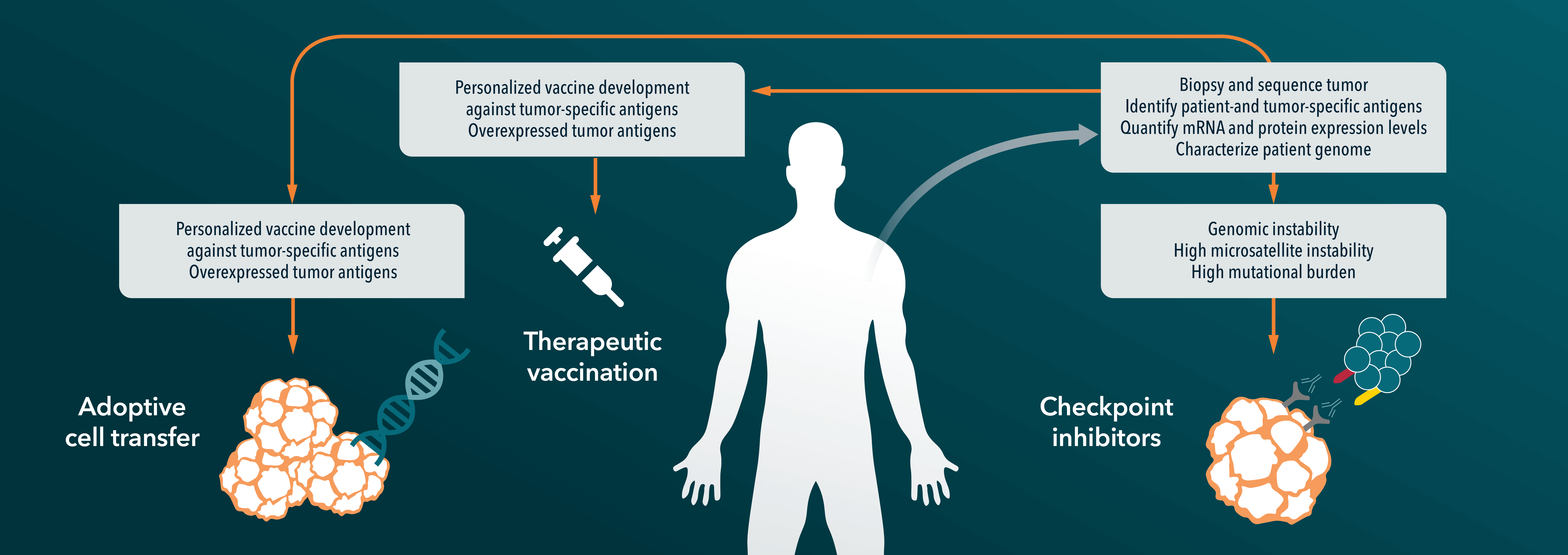
Implementation of digital pathology has also made important contributions to the field of immuno-oncology (I/O). It is now well recognized that the success of immunotherapy depends on the close and dynamic interaction between tumor cells and other cell populations within the TME. The ability to achieve comprehensive spatial analysis of biomarkers with digital pathology enables researchers to better understand the role of this relationship for the development of new and more effective I/O agents.
Technologies for Digital Pathology-Based Biomarker Analysis
A variety of methods are currently used for tissue biomarker detection. These include basic stains that differentiate different cellular compartments (e.g., H&E) or molecular assays such as immunohistochemistry (IHC) or immunofluorescence (IF) for protein biomarkers. Despite advancements in sequencing technologies, methods such as in situ hybridization (ISH) with chromogenic (CSH) or fluorescent (FISH) options remain popular for the detection of RNA/DNA biomarkers.
Since the majority of tissue biomarkers are protein-based, robust IHC and IF protocols have been developed for a number of important cancer targets. These include:
- Tumor associated antigens (TAAs), such as growth factor receptors (e.g., EGFR, HER2)
- Cancer mechanism markers such as tumor proliferation or apoptosis (Ki67, Cleaved Caspase-3), angiogenesis (CD31, VEGF)
- Immune checkpoint inhibitors (ICIs) PD-1 and PD-L1 which can predict response to immunotherapy in some cancer types.
However, chromogenic IHC is limited to a single-marker which renders it unsuitable for modern biomarker studies which often requires detection of multiple markers simultaneously. While serial-section protocols can overcome some of these limitations to a degree, progress in multiplex staining along with advancements in digital pathology imaging and analysis tools have redefined what is now possible for biomarker research.
Detection of multiple protein targets can be accomplished by both IHC and IF, although fluorescent-based assays provide better signal separation which allows for higher complexity and more accurate quantitation.
This is especially important in analyses that rely on very limited tissue samples and can conserve precious samples for other analyses.
The basic principle of multiplexing involves staining of multiple proteins with specific antibodies that are directly or indirectly tagged with distinct chromogens or fluorophores. Depending on the technology and complexity, this can be achieved in a single-step or more commonly, multiple cycles of labeling, imaging, antibody stripping and re-labelling.
Tyramide Signal Amplification (TSA)-based multiplex IHC is one of the most popular methods for the simultaneous detection of multiple proteins. In this system;
- A protein target is labelled with a primary antibody followed by a secondary antibody conjugated with horseradish peroxidase (HRP)
- Incubation with a fluorophore-conjugated tyramide molecule results in the formation of covalent bonds with tyrosine residues on the protein target
- The permanent deposition of fluorophore enables removal of antibodies without loss of fluorescent signal and permits labelling of the next protein of interest.
Importantly, one of the benefits of this approach is that multiple rounds of staining can be carried out using antibodies from the same species. This method is capable of detecting low abundance proteins since fluorophore-conjugated TSA molecules emit a robust signal.
The TSA-based multiplex IHC method has been extensively used for biomarker analysis in I/O applications.
For example, a recent meta-analysis of studies evaluating PD-1/PD-L1 proximity, CD8+ T-cell density in the TME and markers of T cell activation found that multiplex staining was superior to single-staining or other methods (e.g., gene expression profiling) in predicting response to checkpoint inhibitor therapy.
Digital Imaging Platforms for Biomarker Evaluation
There are many commercial and open-source digital analysis platforms available, each with its own set of strengths and weaknesses. ImageJ, CellProfiler, and QuPath are some of the most used open‐source platforms, while HALOTM (by Indica Laboratories) is a professional quantitative pathology option that offers modules for quantification of a variety of biomarker types and supports cellular, tissue, and spatial analysis.
It is highly recommended to consult with experts in digital pathology prior to selecting a tool, so that you have the best chance at selecting the right tool for your specific research questions.
The preparation of high-quality digitized representations of your tissue glass slides is an important factor in developing high quality data. This involves a multistep and complex process that requires strict quality control for each procedure.
Variables to consider when preparing your slides include:
- Fixation protocols and antigen sensitivity
- Tissue processing standard operating procedure (SOP)
- Tissue section thickness
- Staining consistency
- Coverslip material
Lastly, interpretation of chromogenic or fluorescent data from pixels requires an understanding of the settings used to capture, save, and store the images.
This concept also holds true with the use of AI-based algorithms for analysis. Although many image analysis programs come with ‘ready to go’ algorithms/modules, these often need to be adjusted to suit specific research needs.
Conclusion
The opportunity to accelerate cancer drug discovery with digital pathology-based biomarker analysis holds tremendous potential for selecting cancer drug candidates with the most potential for clinical success, including immuno-oncology agents.
The move towards a digital and automated framework reduces the impacts of human error and subjectivity while fostering greater collaboration among researchers for more efficient drug discovery and development.
Crown Bioscience has extensive experience in the use of digital pathology for supporting preclinical biomarker programs for cancer drug discovery and development. Our advanced digital pathology services are designed to help companies accelerate their translational biomarker discovery. Please visit our digital pathology services site to learn more about our expertise in this area.