Preclinical models for prostate cancer serve as essential tools for investigating disease mechanisms, evaluating therapeutic strategies, and supporting the translation of scientific discoveries into clinical applications. Because no single experimental system can fully recapitulate the complexity of this disease, researchers rely on a spectrum of in vitro and in vivo prostate cancer models to address specific scientific and translational questions.
Prostate cancer drug development beyond androgen receptor pathway inhibitors includes protein degraders, radioligand therapies, ADCs, T cell engagers, cell therapies and epigenetic approaches, selecting the right model has become increasingly important (Table 1). Therefore, selecting the right model is not simply a technical decision but a critical component of study design, requiring careful alignment with the biological hypothesis, disease context, and intended clinical application with the therapeutic modality. By ensuring this close alignment between the experimental model and the research and clinical questions being addressed, investigators can enhance the translational impact of their findings.
| Therapeutic Modality |
Critical Model Considerations |
| AR inhibitors/degraders |
AR dependency, AR amplification, AR-V7, CRPC |
| PARP/DDR |
BRCA/HRR status |
| Radioligands |
Target expression, density, heterogeneity, biodistribution |
| ADCs |
Antigen density, internalization, heterogeneity |
| TCEs/BsAbs |
Target expression + human immune system |
| CAR-T/CAR-NK |
Target expression + trafficking/persistence |
| EZH2/epigenetic |
Lineage state, TP53/RB1, NEPC |
| TME therapies |
Immune/stromal composition |
| ICI combinations |
Immune competence and baseline immune phenotype |
Table 1: Overview of Therapeutic Modality and Preclinical Model Considerations
Why Preclinical Models Matter in Prostate Cancer Drug Discovery
Preclinical prostate cancer models allow researchers to investigate key biological processes, including androgen receptor (AR) signaling, genetic and epigenetic changes, interactions between tumors and their surrounding environment, and the mechanisms that drive disease progression and treatment resistance (Table 2). These models are also essential for evaluating new therapies by assessing their ability to slow tumor growth, delay progression, and reduce metastasis.
In addition, preclinical models are critical for understanding why treatments stop working, particularly as prostate cancer evolves into more aggressive forms. They support biomarker discovery by linking molecular features of tumors with treatment responses, helping researchers identify which patients are most likely to benefit from specific therapies. As a result, these models play an important role in advancing personalized treatment strategies and improving the translation of laboratory findings into clinical practice.
| Research question |
Recommended models |
| AR dependence / AR resistance |
Cell lines, organoids, CRPC PDX, castrated/non-castrated mouse models |
| Drug screening |
Cell lines, organoids |
| Tumor heterogeneity |
Organoids, PDX |
| PTEN/PI3K biology |
Genetically characterized PDX, organoids, GEMM |
| DDR/PARP sensitivity |
BRCA/HRR-characterized PDX and organoids |
| Enzalutamide resistance |
Resistant cell lines, organoids, CRPC PDX |
| Lineage plasticity |
Longitudinal organoids, CRPC PDX, GEMM |
| NEPC |
NEPC cell lines, organoids, NEPC PDX, selected GEMMs |
| Bone metastasis |
Bone-tropic models, intratibial/orthotopic approaches, PDX |
| PSMA-targeted therapy |
PSMA-characterized PDX/organoids |
| ADCs |
Antigen-positive PDX + organoids |
| TCE/BsAb |
Humanized models |
| CAR-T/CAR-NK |
Humanized models |
| TME-targeting + ICI |
Syngeneic and/or humanized models |
| Epigenetic therapies |
Lineage-characterized organoids + CRPC/NEPC PDX |
Table 2: Summary of Relevant Models for Specific Research Questions
In Vitro Prostate Cancer Models for Early Drug Discovery
The foundation of this experimental landscape are in vitro prostate cancer models, which include conventional prostate cancer cell lines grown in 2D culture systems (Table 3). These models are widely used because they are accessible, cost-effective, and amenable to high-throughput screening. They provide a simplified and highly controlled system for studying cellular processes, evaluating drug mechanisms of action, and identifying promising compounds. Commonly used prostate cancer cell lines represent different disease states, including androgen-sensitive and androgen-independent phenotypes, allowing researchers to explore key aspects of tumor biology. Unlike other indications, prostate cancer cell line models are limited due to slow growth and dependency on hormonal environment and surrounding TME.
| Cell Line |
AR |
PSA |
Features |
| LNCaP |
++ |
+++ |
AR-mutant |
| C4-2 |
++ |
+++ |
LNCaP derivative; AR-mutant ARv7 |
| 22Rv1 |
++ |
++ |
ARv7 BRCA2 |
| VCaP |
+++ |
++ |
TMPRSS2-ERG fusion, DDR |
| DU145 |
- |
Low |
BRCA2 |
| PC3 |
- |
Low |
BRCA2 |
| PC3-hPSMA |
- |
Low |
Engineered line |
| PC3M-luc |
- |
Low |
Metastatic variant of PC3 engineered to be bioluminescent |
| NCI-H660 |
- |
Neg |
NEPC |
Table 3: Examples of Common Cell Lines for in vitro and in vivo studies
Advanced In Vitro Models to Model the Tumor Microenvironment
3D prostate cancer models, including spheroids and organoids, offer a more biologically relevant alternative. Spheroids are formed by the self-assembly of cancer cells into multicellular structures that better reflect tumor organization, including gradients of oxygen and nutrients. Organoid models go a step further by deriving structures from patient tissue, preserving aspects of tumor architecture, cellular diversity, and genetic heterogeneity.
These prostate cancer organoid models can be expanded and manipulated in culture, making them valuable tools for studying disease biology and drug response in a more patient-relevant context. Organoids can also be derived from patient-derived xenografts (PDXOs) which maintain key characteristics of original PDX (Xu et al. (2023)). A summary of matched prostate PDXOs with PDX are shown in Table 4 along with clinical information.
Co-culture systems further enhance biological relevance by incorporating multiple cell types such as immune cells into the experimental setting. These models enable researchers to examine interactions within the tumor microenvironment, which are critical for understanding processes such as immune evasion, therapy resistance, and metastatic progression. Together, advanced in vitro prostate cancer models provide improved translational value compared with traditional systems, although they may involve greater complexity and lower throughput.
Comprehensive Toolbox of In Vivo Prostate Cancer Models
In vivo prostate cancer models complement in vitro approaches by capturing the systemic and microenvironmental factors that influence tumor behavior in living organisms. Among the most widely used are cell line-derived xenograft (CDX) models, in which established prostate cancer cell lines are implanted into immunocompromised mice (Table 3). CDX systems allow for relatively rapid and reproducible evaluation of tumor growth and drug response, making them useful for early-stage efficacy testing, mechanism of action studies, resistance engineering and orthotopic/metastatic modeling as well as incorporation into humanized mouse models for immune-oncology and combination studies (Table 2).
For dissecting AR mechanism, in vivo studies can be conducted in either castrated or non-castrated mice. The 22Rv-1 CDX model, derived from the xenograft line CWR22R from a patient with bone metastasis, is AR+ but only partially sensitive to testosterone due to expression of ARv7 splice variant, thus able to grow in castrated mice and poor response to both Enzalutamide and Abiraterone as well as Docetaxel (Figure 1).
Due to this resistant profile, 22Rv1 is an ideal model to evaluate novel agents to overcome resistance that is driven by the ARv7 splice variant. VNPP433-3b, a next generation galeterone analog (NGGA) molecular glue degrader that targets AR, including splice variants, has been reported to significantly reduce TGI in 22Rv1 tumors compared to Enzalutamide in a head to head comparison (Kwegyir-Afful et al. (Cancers, 2019).
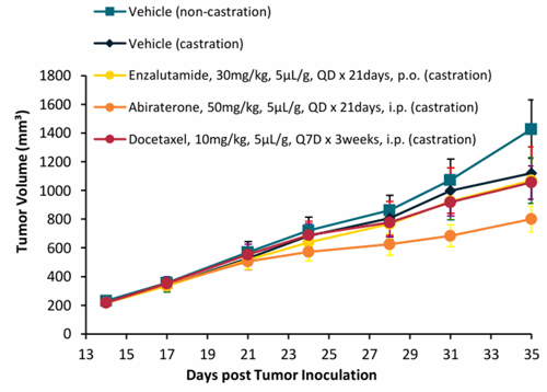 Figure 1: In vivo CDX 22Rv1 (ARv7 positive cell line) response to castration, Enzalutamide, Abiraterone and Docetaxel
Orthotopic prostate cancer models, in which tumor cells are implanted into the prostate of the host animal, offer the advantage of placing tumors in their anatomically relevant environment. As a result, they allow for more realistic interactions with surrounding tissues and can influence tumor growth patterns and metastatic spread.
Metastatic prostate cancer models, designed to replicate the dissemination of cancer cells to distant sites such as bone, are especially important given the clinical significance of bone metastasis in advanced disease. While bone metastasis models remain challenging to establish and standardize, they are critical for understanding disease progression and evaluating therapies aimed at late-stage cancer.
Patient-derived xenograft (PDX) models represent a more advanced category of prostate cancer xenograft models. In these systems, tumor tissue, either from primary tumor or metastatic site, obtained directly from patients is implanted into mice, and unlike cell line models PDXs do not adapt to 2D culture due to maintenance in vivo, preserving many aspects of the original tumor, including its histology, genetic profile, and heterogeneity (Xie et al. (JCI Insight, (2026). A summary of the prostate PDX models with matched organoids, patient information and AR status are shown in Table 4.
Prostate cancer PDX models are particularly valuable for translational research because they can better reflect clinical responses and resistance mechanisms. When combined with clinical annotation, these models can also support biomarker development and the identification of patient subgroups. When matched with in vitro organoids, more extensive drug screening, MoA studies and resistance profiling as well as drug combination studies can be performed before in vivo studies. Figure 2 shows two PDX models derived from mCRPC with high levels of AR, TMPRS-ERG fusion and PTEN loss (Table 4).
PR9585, which was experimentally derived to select out resistant subclones was unresponsive to castration, Docetaxel and Enzalutamide, whereas PR9587 was sensitive to castration suggesting some potential heterogeneity in PR9587. In vitro dose-response curves showed similar resistance in the organoid derived from PR9585 (Figure 2C) to Enzalutamide and Docetaxel as well as Abiraterone and Cabazitaxel.
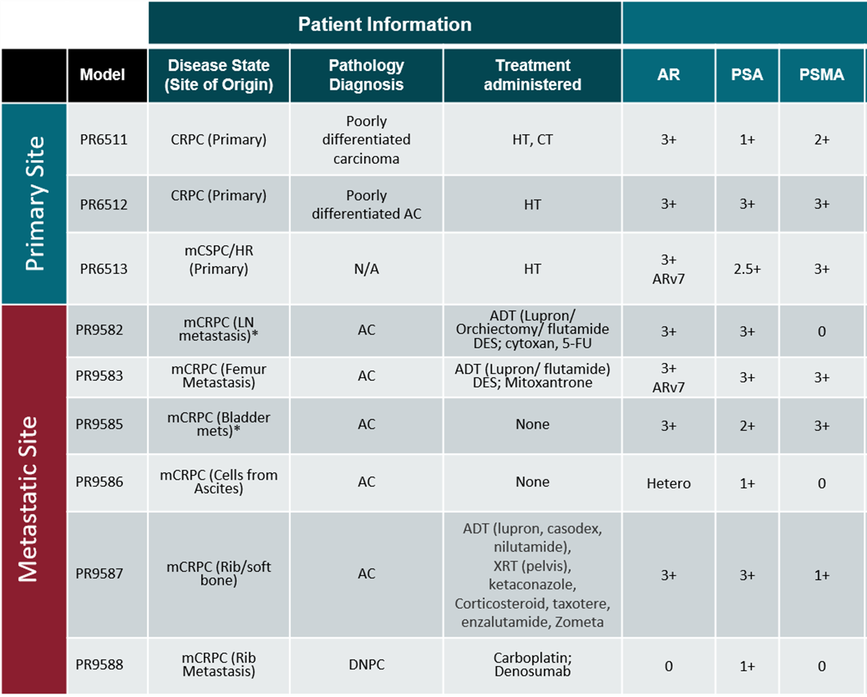 Table 4: Summary of Prostate Cancer PDX Models and Matched PDXO
(a)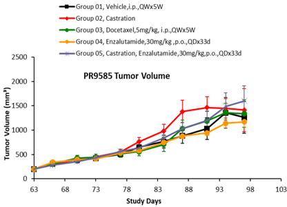 (b)
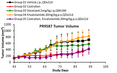
Figure 2: In Vivo Response of CRPC AR+ PDX Models PR9585 (A) and PR9587 (B) to Castration and Treatment with Enzalutamide and Docetazel and In Vitro CTG Readout in Matched PDXO PR9585B
As mentioned in previous blog posts the characterization of models should encompass expression for key markers and tumor associated antigens (TAA) such as AR, PSA, PSMA, STEAP1, B7H3, KLK2 and other NE markers, genetic features (AR mutation or splice variants, BRCA1/2, PTEN, TP53, RB1 loss) and response to standard of care to determine responder/non-responder profiles. High concordance of tumor associated antigen expression was reported between PDX and their matched PDXO (AACR 2026 Poster 7540) as well as proteomics (AACR 2026 Poster 7679) and molecular similarity to patient tumors (Xie et al., JCI Insight, (2026).
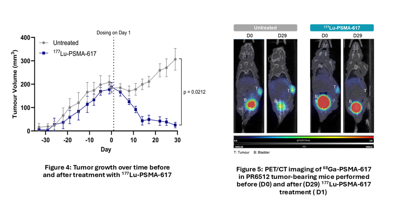
Targeting PSMA in prostate cancer has led the progress in radiopharmaceuticals such as Pluvicto, a Lutetium-177 radioactive payload targeting PSMA for mCRPC. End-to-end in vitro and in vivo evaluation of radiopharmaceuticals requires specialized expertise in radiochemistry integrated with preclinical modeling (AACR 2026 Poster 7370). 177Lu-PSMA-617 demonstrated PSMA targeted tumor reduction in 2D and 3D spheroid and organoids (Figure 3) as well as in vivo PDX models of mCRPC (Figure 4). Theranostic pairing with 68GA-PSMA-617 PET-CT imaging tumor burden could be visualized and quantified, showing clearance via the bladder and reduction in tumor after treatment (Figure 5).
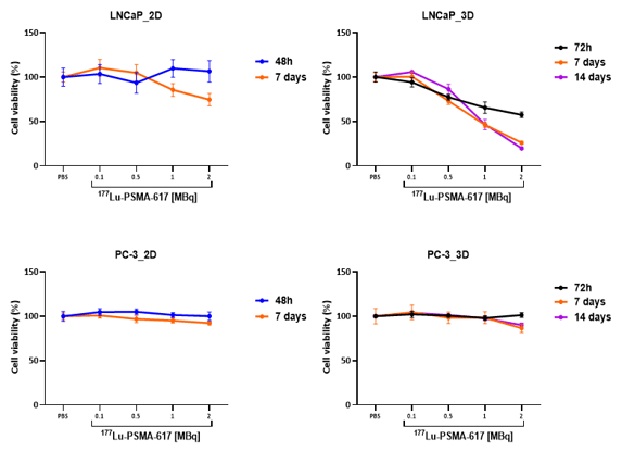
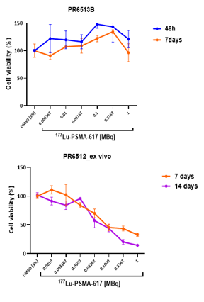
Figure 3: Cells in either 2D or 3D spheroid cultures were exposed for 3h to the lutetium labelled therapy and CTG readout was taken 2/7 days after treatment. LNCaP (high PSMA) showed sensitivity to the treatment in a dose dependent manner and no effect in PC-3 (PSMA negative).
Immuno-Oncology Models: Evaluating the Tumor–Immune Interface in Prostate Cancer
Immunotherapy presents a particular challenge in prostate cancer drug development. Unlike many highly immunogenic tumor types, prostate cancer is generally considered an immunologically cold or immune-suppressed tumor, characterized in many cases by limited T cell infiltration, impaired antigen presentation and an immunosuppressive tumor microenvironment (TME). These features can contribute to the limited clinical activity of immune checkpoint inhibitors in unselected patient populations and highlight the need for models that capture the interactions between tumor cells, immune populations and the surrounding stroma.
The emergence of new immunotherapeutic approaches, including bispecific antibodies and T cell engagers (TCEs), CAR-T and CAR-NK cell therapies, and strategies designed to remodel the TME, creates additional requirements for preclinical model selection (Table 5). The most appropriate model depends on the mechanism of action being investigated and the specific biological question, including target expression, immune-cell recruitment, tumor infiltration, immune activation and the development of resistance.
Syngeneic models such as RM-1 provide an immunocompetent setting for investigating immune checkpoint inhibitors, immune-modulating agents and combinations designed to overcome immune suppression. Key endpoints include CD8+ and CD4+ T cell infiltration, regulatory T cells, macrophages, myeloid-derived suppressor cells (MDSCs), dendritic cells and cytokine profiles.
Humanized PDX models combine patient-derived tumors with a reconstituted human immune system, enabling evaluation of selected human-specific immunotherapies, including T cell engagers, bispecific antibodies, checkpoint inhibitors and cell therapies.
PBMC-based PDX/CDX models offer relatively rapid T cell reconstitution but are limited by alloreactivity and graft-versus-host disease (GvHD), while CD34+ stem cell-based models can provide broader immune-cell reconstitution, although immune maturation and functionality may remain incomplete. Careful qualification of immune engraftment, composition and function is therefore essential.
Genetically engineered mouse models (GEMMs) enable the study of prostate cancer initiation and progression in an immunocompetent setting e.g TRAMP-C2 model. These models can recapitulate specific genetic alterations observed in patients and allow for longitudinal analysis of tumor development. Although they can be resource-intensive and less flexible than transplant models, they are valuable for studying tumor biology within the context of an intact immune system.
Development of homograft models, where tumor from GEMM is implanted into donor mice for expansions, can streamline this process. In addition generation of cells over expressing relevant TAA such as PSMA can also facilitate evaluation of human targeting agents in combination with immunotherapeutics.
| Therapeutic approach |
Key biological questions |
Suitable models |
| Immune checkpoint inhibitors and TME-modulating agents |
Immune activation, infiltration and reversal of immune suppression |
Syngeneic models; selected humanized PDXs |
| T-cell engagers and bispecific antibodies |
Target expression, T-cell recruitment and tumor-cell killing |
Humanized PDXs; organoid–immune-cell co-cultures |
| CAR-T and CAR-NK therapies |
Target recognition, tumor infiltration and immune-cell persistence |
Humanized PDXs; organoid–immune-cell co-cultures; selected syngeneic surrogate system |
Table 5: Matching immunotherapeutic approaches to preclinical models
Integrated Biomarker Analysis
For immunotherapeutic programs, tumor growth inhibition alone is insufficient to establish mechanism of action. Evaluation should integrate efficacy with immune profiling, including immune-cell composition, activation and function, cytokine production, target expression and spatial distribution of immune cells within tumor tissue. Techniques such as flow cytometry, multiplex immunohistochemistry, immunofluorescence and spatial profiling can help identify treatment-induced changes and potential resistance mechanisms.
Ultimately, combining immunocompetent models, humanized PDXs and patient-derived tumor–immune-cell co-cultures provides complementary approaches to evaluating immunotherapies. Model selection should be driven by the therapeutic mechanism and the specific tumor–immune interactions that need to be investigated.
Specialized Expertise to Navigate the Limitations of Current Prostate Cancer Models
While modern in vitro and in vivo systems have improved researchers’ ability to study disease biology and evaluate therapeutic candidates, no model can fully capture the complexity of human prostate cancer. Understanding these limitations is essential for designing effective drug discovery strategies.
One of the most significant challenges is the inability of many models to capture the full heterogeneity of patient tumors.
Studies focused on biomarker discovery, patient stratification, drug sensitivity testing, or prediction of clinical response often require models that closely resemble human tumors at the molecular and phenotypic levels. Aligning model choice with the desired translational endpoint helps ensure that preclinical findings generate actionable data for future clinical development. Prostate cancer is a highly diverse disease, with significant differences in its molecular and biological characteristics both between patients and within individual tumors. As a result, findings from a single model may not apply broadly, making it important to confirm results using patient derived models such as PDXs.
Low establishment efficiency and limited availability of suitable tumor material
Primary prostate tumors can be difficult to establish in culture, particularly from localized disease. Metastatic biopsies may provide better material for advanced disease modeling, but sample availability and tumor content remain limiting factors. Access to a broad range of models, tissue sourcing and application of relevant model systems and expertise is an essential component of prostate cancer drug discovery.
Modeling metastasis remains a particularly difficult challenge.
Bone is the most common site of metastasis in advanced prostate cancer; however, the bone microenvironment is highly complex. Traditional cell culture systems cannot fully reproduce this complexity, and many animal models only partly reflect how bone metastases develop in patients. Although, accurately testing therapies designed to target prostate cancer in bone remains challenging, advanced experimental models of metastasis combined with imaging, such as bioluminescent optical imaging or ultrasound, has enabled monitoring of disease that reflects later stages of prostate cancer (Table 2).
Studying hormone dependency and independency can be complex. Prostate cancer progresses through several biological stages, from disease that depends on AR signaling to more advanced forms that can grow despite AR-targeted therapies. Studies of hormone-sensitive prostate cancer require models that maintain functional AR activity and respond to hormonal changes. Different models have different levels of androgen dependence, making standardized conditions difficult to apply across a panel.
For example androgen-dependent models may require testosterone supplementation to support tumor establishment and growth. Castration or androgen deprivation can alter tumor growth and select for castration-resistant populations. For example, VCaP and LNCaP models retain important features of AR-driven prostate cancer biology, but their growth characteristics and androgen requirements differ. Any changes in androgen availability can influence the interpretation of treatment efficacy, particularly for AR-targeted therapies so pilot studies are needed to assess the growth dependency of prostate cancer to determine optimal conditions (Figure 1).
In contrast, research on castration-resistant prostate cancer (CRPC) and AR-independent disease relies on models that capture the mechanisms of treatment resistance, disease progression, and changes in tumor cell identity. Treatment-induced lineage plasticity, including the transition from adenocarcinoma to neuroendocrine prostate cancer (NEPC), can be difficult to reproduce in standard culture systems. Expansion of PDX and organoid models with characterization of hormone dependency continues to provide opportunities for a wider selection of novel models, heterogeneity and mechanisms of resistance beyond the current cell line prostate cancer panel.
Reproducing treatment resistance and disease progression
Advanced prostate cancer is not a single biological entity. Tumors can evolve under treatment pressure through several mechanisms, including:
- AR amplification, mutation and splice variants such as AR-V7.
- Activation of alternative signaling pathways.
- Loss of tumor suppressors such as PTEN, TP53 and RB1.
- Lineage plasticity and transformation towards neuroendocrine or other AR-independent phenotypes.
- Changes in cell-surface antigen expression, potentially affecting ADCs, radioligands and T cell-directed therapies
Capturing these dynamic changes requires models that reflect the relevant disease stage and treatment history. A model derived from untreated disease may not adequately represent heavily pretreated metastatic castration-resistant prostate cancer (mCRPC) thus selecting models that mirror the clinical disease requires well-characterized models with relevant benchmark data.
Practical considerations can also limit the use of more sophisticated model systems.
Advanced platforms, such as organoids, co-cultures, orthotopic systems, PDX or humanized models, often require specialized expertise, lengthy development timelines, and substantial financial investment. These factors can restrict their accessibility and make large-scale studies difficult to perform. In contrast, simpler cell-based assays provide higher throughput and lower costs but may sacrifice biological relevance.
Many models cannot accurately reproduce long-term disease progression and the emergence of treatment resistance.
Many preclinical models are optimized to evaluate short-term responses to therapeutic interventions, but prostate cancer often progresses over years through a series of adaptive molecular changes. Replicating this evolutionary process in experimental systems remains challenging, making it difficult to accurately predict mechanisms of acquired resistance, lineage plasticity, or progression to aggressive disease states.
Our 4 step strategy to resistance modeling provides guidance on understanding and characterizing the resistant phenotype and finding the right model to match the profile such as clinically pretreated patient models, induced models of resistance or engineered specialized models
Ultimately, successful prostate cancer drug discovery often relies on a combination of models. Using multiple complementary systems helps researchers confirm findings in different settings, strengthen confidence in preclinical results, and improve the chances that promising therapies will translate successfully to patients.
Our teams of specialist scientists are experienced in delivering both standard and complex in vitro and in vivo systems and can therefore provide guidance on identifying the right models to support any drug program. Together, these translational strategies help maximize the strengths of existing prostate cancer models while mitigating their limitation
Conclusion
Preclinical prostate cancer models are indispensable tools for understanding disease biology and advancing drug discovery, but their value depends on thoughtful selection and strategic use. By combining different platforms, incorporating molecular data, and focusing on clinical relevance, researchers can maximize the impact of their studies and generate insights that translate more effectively to patient care. The key question is therefore not simply which prostate cancer model is best, but which combination of models provides the most relevant evidence for the biological and therapeutic question being asked.
References
Peer-Reviewed Journal Articles
- Xu, X., et al. (2023). A living biobank of matched pairs of patient-derived xenografts and organoids for cancer pharmacology. PLoS ONE, 18(1), e0279821. PLOS ONE Journal Portal
- Kwegyir-Afful, A. K., et al. (2019). Galeterone and The Next Generation Galeterone Analogs, VNPP414 and VNPP433-3β Exert Potent Therapeutic Effects in Castration-/Drug-Resistant Prostate Cancer Preclinical Models In Vitro and In Vivo. Cancers, 11(11), 1637. MDPI Open Access Platform [1]
- Xie, Z., et al. (2026). The molecular similarity landscape of preclinical cancer models to patient tumors. JCI Insight, 11(16), e206449. JCI Insight Journal Portal
Conference Abstracts and Posters
- Tu, X., et al. (2026). Integrated Multi-omics Analysis Reveals Conserved Tumor-Associated Antigens (TAAs) Profiles in PDX and Organoid Models for Advancing ADC Development [Poster 7540]. AACR Annual Meeting 2026. Crown Bioscience Poster Library
- Mao, B., et al. (2026). Advanced Proteomic Profiling of Patient-derived Tumor Models Reveals High Cross-model Concordance and Improved Translational Fidelity [Poster 7679]. AACR Annual Meeting 2026. Crown Bioscience Poster Library
- Liu, A., et al. (2026). Medicines Discovery Catapult Collaboration: End-to-end In Vitro and In Vivo Evaluation of Radiopharmaceuticals in Patient-derived Models [Poster 7370]. AACR Annual Meeting 2026. Crown Bioscience Poster Library