Key assays and analytical techniques for the development of antibody drug conjugates
10:46
SHARE
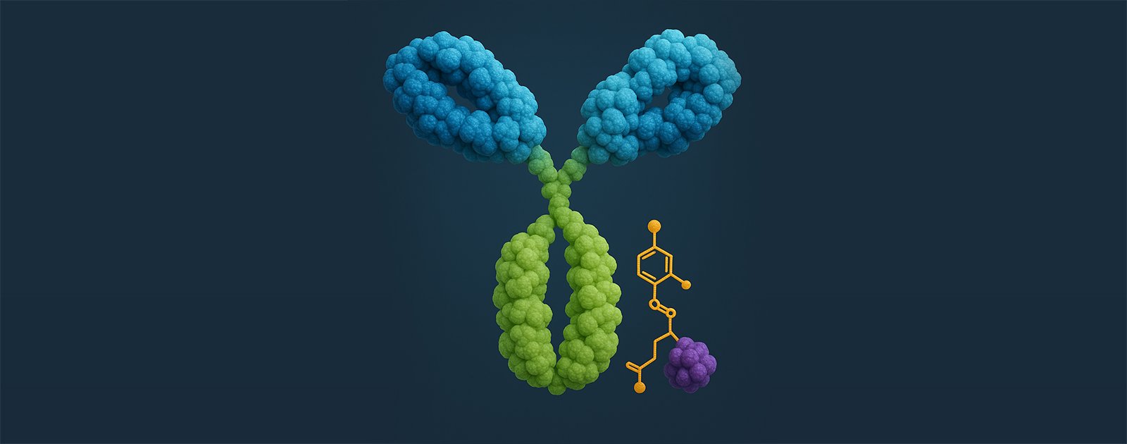
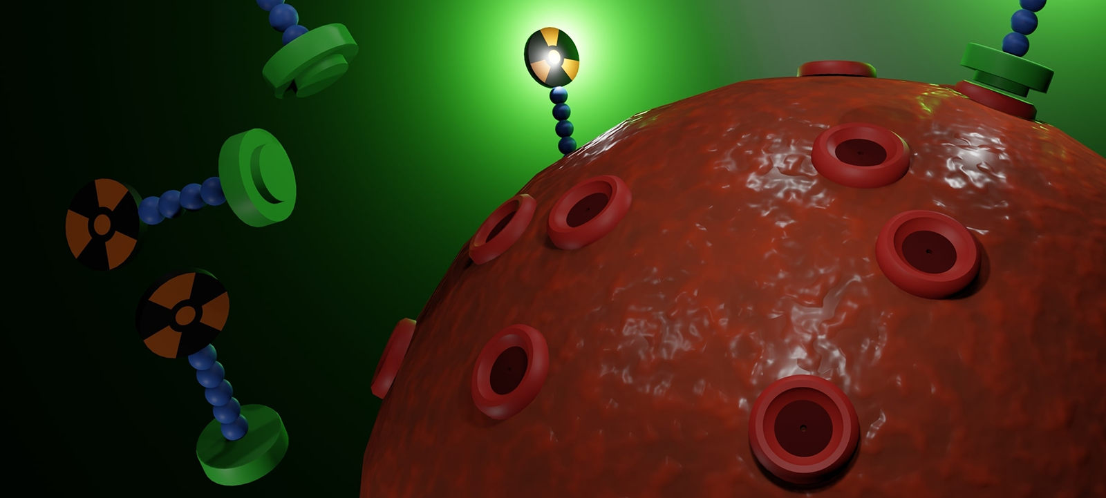
Highly targeted antibody-drug conjugates (ADCs) combine an antibody with a cytotoxic payload, covalently attached via a chemical linker. These “biological missiles” represent an exciting new advance in anti-cancer therapies and are one of the fastest-growing segments in oncology drug development. Due to their complex make-up and multiple components, analysis of their chemistry and functionality is challenging, requiring advanced assays and techniques for effective study and characterization. This article introduces key assays and models available to research teams looking to investigate ADC mechanisms and develop the next generation of this exciting new drug category.
Key assays for ADC development
Ligand-binding assay (LBA)
An LBA is biochemical test that measures the interaction between a ligand (such as a small molecule) and a biomolecular target (for ADCs, an antibody). They can be used to determine if an ADC can attach to cancer cells and they can help verify the consistency and effectiveness on an ADC based on how many drug molecules are attached to each antibody. Total antibody (TAb) or total conjugated ADC can be measured by using agents that bind either to the antibody or payload within the ADC before antibodies are introduced that detect the presence of antibodies on the ADC itself.
LBAs also reveal information about the way ADCs behave in the body, namely how long they stay in the bloodstream of a patient and if antibodies working against the ADC (which could reduce their effectiveness) are created in the patient’s body. In some cases, cytotoxic payloads are structurally altered by metabolic processes. In these instances, it is possible to use to an LBA to measure just the active ADC payloads.
The LBA has long been the favored bioanalysis platform for ADC measurements due to its sensitivity, precision and high sample throughput. Additionally, they are generally considered lower-cost and more straightforward than other assays available. They are particularly valuable for research teams focused on large molecules or anti-drug antibodies.
UV-Vis spectrophotometry: Relatively quick and simple but cannot offer information about drug load distribution. Less accurate than other methods, providing an estimate of DAR only.
Cathepsin-B enzyme-based digestion: More complex than UV-Vis spectrophotometry but can overcome some of its limitations. Cannot offer information about drug load distribution.
LC-MS: Suitable for detailed DAR analysis, it can also be used to assess the drug load distribution of different small molecular drugs to identify different ADC forms.
Reversed-phase liquid chromatography (RP-HPLC): Suitable for more detailed DAR analysis and drug load distribution evaluation, including small molecular drugs at light- and heavy-chain levels.
Hydrophobic interaction chromatography (HIC): The most widely used method for more detailed DAR analysis and drug load distribution evaluation. Particularly compatible with cysteine-linked ADCs.
Additionally, it’s essential to understand how the DAR may change after an ADC enters the body and drug molecules detach or transform. If this happens, the DAR value will no longer match the reference sample used for measurement, meaning any test results won’t be accurate to clinical settings. Case studies have shown the implications of this for research terms and the implications for patients, reiterating the importance of “DAR-insensitive” tests.
Organoids are emerging are a key tool in the fight against resistance. Along with ex vivo cultures, they have proven to be the most effective models for identifying specific resistance mechanisms. As their three-dimensional structures mimic the architecture and functionality of tumor structures, they are particularly appropriate for testing individual ADC components and mechanisms.
Organoid model banks, like those held by Crown Bioscience allow research teams to access fully characterized preclinical models that recapitulate specific resistance phenotypes or create appropriate models (via drug-challenge, CRISPR engineering, transduction, and other technical advances) where relevant models don’t exist. For example, Crown Bioscience holds more than 200 patient-derived organoids and over 500 patient-derived xenograft (PDX)-derived organoids, including 40+ models pretreated with ADC therapeutics.
These organoid models support high-throughput studies, allowing developers to test multiple drug combinations, dosing schedules, and ADC variants against the same resistance tissue. The OrganoidXploreTM platform reduces study timelines to six weeks to reduce the overall development pipeline and increase the chances of successfully developing novel ADCs or drug combinations or identifying key resistance biomarkers.
Investigating ADC linker stability
The linker in an ADC provides a bridge between the cytotoxic drug and the antibody. Linker stability is crucial for ADC efficacy as it ensures that the payload is only released once it is inside the cancer cell rather than too early, which risks releasing the drug systemically and increasing side effects. Additionally, linker stability helps to keep ADCs intact in the bloodstream for longer and can enhance the “bystander killing effect”, where ADCs kill adjacent, antigen-negative cancer cells.
In vivo models, like PDXs, mimic the clinical setting. This means they can be used to investigate linker stability as they allow researchers to test hypotheses developed in vitro, analyze the efficacy of drugs in a physiological setting, and analyze the toxicity and metabolism of the ADC. Crown Bioscience offers the world’s largest collection of highly characterized PDX models, including over 300 pretreated models covering 25 cancer types derived from patient on different drug treatments.
Immune responses
The primary mechanism of an ADC involves delivering a toxic payload directly into a target cell, but the antibody within the ADC can also be used to trigger immune responses to kill cancer cells. The immune responses that can be utilized to optimize ADCs include antibody-dependent cellular cytotoxicity (ADCC), where the antibody in the ADC binds to the cancer cell and attracts immune cells which kill the cancer cell, and antibody-dependent cellular phagocytosis (ADCP), where the antibody attaches to the cancer cells, triggering macrophages to engulf these target cells.
However, ADCs also have the ability to provoke immunogenicity (unwanted immune responses in patients) when the body produces anti-drug antibodies, reducing the efficacy of the ADC. In this instance, ADCC and ADCP can be triggered against the ADC rather than enhancing its action. This can cause the body’s immune system to work against the ADC, clearing it from circulation more quickly and neutralizing its therapeutic action.
Investigating how these two processes can be enhanced or mitigated requires a variety of pre-clinical model types. This may include:
Co-culture systems that combine cancer cells with macrophages (or other immune effector cells) so immune-mediated killing can be observed
Organoid models that reveal cell-cell interactions in a more physiologically relevant environment
PDX in humanized mice that reveal more complete immune interactions and reflect patient tumor heterogeneity
Due to the complexity of ADCs, no one model can provide a complete picture of their functionality. In vitro models can be utilized during earlier pre-clinical stages for controlled mechanistic studies, while in vivo models can be used to validate hypotheses and give insight into physiological contexts. When employed strategically, the inherent strengths of each model can be maximized at the right stage to help build confidence and increase the chances of success.
Conclusion
ADCs represent a promising new avenue in oncology treatment. Their complex make-up and mechanisms are being matched with ever-advancing assays, techniques, and models to help research teams overcome resistance, optimize ADC structures and linker stability, and utilize immune responses to maximize the potential of this new technology.
Navigate the Complexities of ADC Development
Cite this Article
Wilkin, B., (2025) Key assays and analytical techniques for the development of antibody drug conjugates - Crown Bioscience. https://blog.crownbio.com/key-assays-and-analytical-techniques-for-the-development-of-antibody-drug-conjugates
Antibody-drug conjugates (ADCs) are among the most exciting advances in target cancer therapy. They combine the specificity of monoclonal antibodies w…
Antibody-drug conjugates (ADCs) represent a cutting-edge advance in cancer therapy. These targeted agents combine a monoclonal antibody and a cytotoxi…
Introduction to Theranostics: Bridging Imaging and Therapy The convergence of diagnostics and therapeutics—collectively termed theranostics—is revolut…