

Harnessing Mass Spectrometry for Biomarker Discovery in AML Drug Development
August 7, 2025

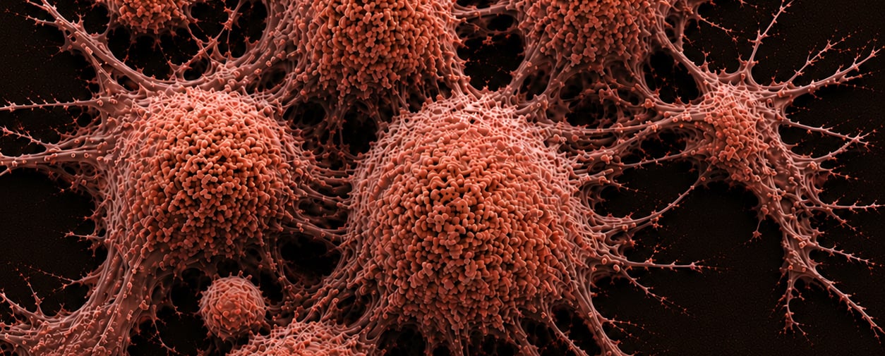
Acute myeloid leukemia (AML) remains one of the most aggressive hematological malignancies, characterized by the rapid proliferation of immature myeloid cells in the bone marrow and blood. Despite advances in chemotherapy and hematopoietic stem cell transplantation, AML continues to pose significant clinical challenges due to its high relapse rate, clonal heterogeneity, and resistance to standard therapies. The five-year survival rate remains low, especially among older adults, underscoring the urgent need for more personalized and effective treatment strategies. In this context, precision medicine—guided by robust biomarkers—is emerging as a transformative approach to improve outcomes in AML drug discovery.
The drive toward precision oncology in AML has intensified the search for clinically actionable biomarkers that can aid in early diagnosis, risk stratification, therapeutic targeting, and real-time monitoring of treatment response. However, identifying such biomarkers in AML is particularly complex due to the dynamic nature of the disease, inter-patient variability, and the heterogeneity of subclonal populations. Traditional biomarker discovery methods, while useful, often fall short in capturing the full molecular landscape of AML. This has led researchers to adopt more comprehensive, high-throughput technologies capable of interrogating multiple biomolecular layers simultaneously.
Among these, mass spectrometry (MS) has established itself as a powerful and versatile platform for AML biomarker discovery and validation. Its ability to perform deep proteomic, metabolomic, and lipidomic profiling with high sensitivity and specificity makes it uniquely suited for uncovering disease-associated molecular signatures in AML. By enabling the detection of low-abundance analytes, post-translational modifications, and metabolic shifts, MS is unlocking new insights into AML pathogenesis and therapeutic vulnerabilities. Moreover, as mass spectrometry workflows become more streamlined and clinically compatible, their integration into drug development pipelines is accelerating the identification of novel targets and predictive biomarkers.
This article explores how biomarker and mass spectrometry technologies are converging to accelerate the development of more effective AML therapies. We will discuss how MS platforms facilitate the discovery of diagnostic, prognostic, and predictive biomarkers, the workflows used to validate them, and the clinical implications of these advancements for future AML treatment paradigms.
Introduction: The Need for Precision in AML
AML is a genetically heterogeneous disease with complex molecular alterations that include mutations in genes such as FLT3, NPM1, IDH1/2, and TP53. These genetic abnormalities drive distinct biological behaviors and clinical outcomes, making AML notoriously difficult to treat with a one-size-fits-all approach. Despite remarkable progress in genomics and the development of targeted agents like FLT3 and IDH inhibitors, the 5-year survival rate for AML remains below 30% in adults, largely due to the lack of robust biomarkers that can inform treatment decisions throughout the disease course. This gap underscores the need for dynamic, multi-dimensional biomarker platforms that go beyond genomics to capture the full molecular complexity of AML.
Biomarkers, when effectively identified and validated, can significantly improve therapeutic outcomes by enabling:
- Early diagnosis and risk stratification to tailor induction strategies
- Real-time assessment of drug efficacy during clinical trials and treatment
- Patient-specific therapeutic targeting through companion diagnostics
- Monitoring of minimal residual disease (MRD) to detect early relapse and guide post-remission therapy
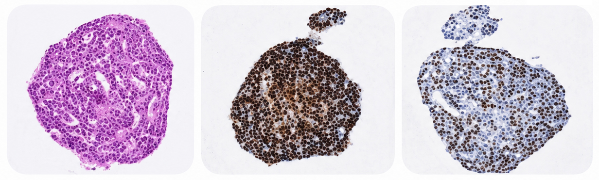
However, the path to actionable biomarker discovery in AML is fraught with challenges. The biological complexity of AML, which includes the coexistence of multiple subclones and dynamic shifts in mutational landscapes, complicates the identification of consistent markers across patients. Additionally, sample variability—due to differences in specimen type (bone marrow vs. peripheral blood), disease stage, or processing methods—can introduce noise and obscure true biological signals. Traditional platforms like ELISA and immunohistochemistry are limited by their reliance on antibodies, which may lack the sensitivity or specificity needed to detect low-abundance proteins or subtle post-translational modifications.
This is where mass spectrometry (MS) has emerged as a game-changing technology. With its ability to perform unbiased, high-throughput analysis of complex biological samples, MS enables comprehensive profiling of proteins, metabolites, and lipids associated with AML disease progression, therapeutic response, and relapse. Unlike traditional methods, MS does not require prior knowledge of the target and can simultaneously analyze thousands of biomolecules with exquisite sensitivity. This makes it an ideal platform for uncovering novel biomarkers that might otherwise go undetected and paving the way for more effective, individualized therapies in AML.
The Power of Mass Spectrometry in Biomarker Research
Why Mass Spectrometry?
Mass spectrometry (MS) is a highly sensitive analytical technique that identifies and quantifies molecules based on their mass-to-charge ratio. It offers several critical advantages over conventional biomarker platforms, including:
- High sensitivity and specificity for detecting low-abundance proteins, peptides, lipids, and metabolites
- Label-free quantification, eliminating the need for antibodies or fluorescent tags
- Multiplexing capabilities to simultaneously analyze thousands of biomolecules in a single run
- Versatility across diverse biological samples, including plasma, bone marrow, urine, cerebrospinal fluid, and cell lysates
In the context of AML, these features enable MS-based platforms to overcome key limitations of traditional antibody-based assays, such as cross-reactivity, limited dynamic range, and dependence on known targets. Mass spectrometry allows for unbiased discovery, offering a more comprehensive and high-resolution view of the molecular landscape in AML patients.
Furthermore, MS excels in detecting post-translational modifications (PTMs)—such as phosphorylation, acetylation, ubiquitination, and glycosylation—which are often critical in cancer biology but difficult to measure with conventional methods. These PTMs can reveal dysregulated signaling pathways and therapeutic vulnerabilities in AML that are not captured by DNA or RNA sequencing. For example, phosphoproteomics can uncover aberrant kinase activity, enabling the development of kinase inhibitors or combination therapies tailored to the patient's disease profile.
Another major strength of mass spectrometry lies in its quantitative reproducibility, making it suitable not only for discovery-phase research but also for biomarker validation and clinical translation. Recent advances in targeted MS techniques, including Multiple Reaction Monitoring (MRM) and Parallel Reaction Monitoring (PRM), have made it possible to develop highly precise assays for quantifying candidate biomarkers in large patient cohorts. These assays are increasingly being used in early-phase clinical trials to stratify patients, monitor pharmacodynamic responses, and evaluate therapeutic efficacy—bringing mass spectrometry closer to routine clinical use in AML management.
Workflow: Using Mass Spectrometry to Discover and Validate Biomarkers in AML
1. Sample Preparation and Enrichment
AML biomarker discovery often begins with sample collection from bone marrow aspirates or peripheral blood, the two primary sources of disease-relevant material. Due to the inherent complexity and variability of biological matrices, rigorous sample preparation is essential to ensure reproducibility and sensitivity. This may involve:
- Depletion of high-abundance proteins (e.g., albumin, immunoglobulins) that can mask low-abundance targets
- Enrichment techniques to isolate rare proteins or post-translational modifications (PTMs) such as phosphorylation or glycosylation
- Fractionation using liquid chromatography or capillary electrophoresis to reduce sample complexity
These preprocessing steps are vital for maximizing signal-to-noise ratio and improving the depth and reliability of mass spectrometry analyses, particularly in low-input or heterogenous AML samples.
2. Discovery Phase: Untargeted Proteomics and Metabolomics
In the discovery phase, untargeted or "shotgun" mass spectrometry is employed to explore the full spectrum of biomolecules present in a sample without prior assumptions. This approach enables high-throughput identification of differentially expressed proteins, peptides, and metabolites between patient subgroups—such as responders vs. non-responders, or relapsed vs. treatment-naïve individuals.
- LC-MS/MS (Liquid Chromatography–Tandem Mass Spectrometry) is widely used to generate high-resolution proteomic profiles.
- Metabolomic profiling through MS uncovers pathway-level metabolic alterations that may serve as therapeutic vulnerabilities or early indicators of drug resistance.
- Label-free quantification and isobaric tagging methods like Tandem Mass Tags (TMT) and Isobaric Tags for Relative and Absolute Quantitation (iTRAQ) facilitate accurate cross-sample comparisons, essential for biomarker discovery in multi-patient cohorts.
This phase is exploratory, aiming to build a comprehensive inventory of potential biomarkers for downstream validation.
3. Bioinformatics and Statistical Analysis
The high-dimensional datasets generated by MS require advanced bioinformatics pipelines for proper interpretation. Key steps include:
- Data normalization to correct for technical variability
- Batch effect correction to ensure consistency across runs
- Differential expression analysis to highlight statistically significant molecular changes
- Pathway enrichment analysis to identify biologically relevant mechanisms
- Network modeling to explore functional interactions between biomolecules
Integrating proteomic and metabolomic data with clinical metadata (e.g., cytogenetics, response to therapy, survival outcomes) and genomic profiles (e.g., mutational status) allows for a systems biology approach to biomarker prioritization. This generates a curated list of candidate biomarkers with diagnostic, prognostic, or therapeutic significance.
4. Validation Phase: Targeted Mass Spectrometry
Following the discovery phase, shortlisted biomarkers undergo rigorous validation using targeted MS techniques such as Selected Reaction Monitoring (SRM) or Parallel Reaction Monitoring (PRM). These assays are highly quantitative, offering:
- High precision and reproducibility, essential for clinical relevance
- Low limits of detection, suitable for low-abundance biomarkers in complex biological samples
- Use of stable isotope-labeled internal standards for absolute quantification and assay standardization
Validated biomarkers can then be transitioned into clinical assay development, conforming to Good Laboratory Practice (GLP) and regulatory requirements. In the context of AML, validated MS-based assays are increasingly being integrated into early-phase trials and companion diagnostics, where they support patient stratification, real-time monitoring of drug response, and assessment of minimal residual disease (MRD).
Importantly, this workflow is iterative and scalable. As new data emerges, previously validated biomarkers can be refined, expanded, or combined into multi-marker panels, enhancing their predictive power and clinical utility.
Case Studies: Applications of MS-Based Biomarker Discovery in AML
1. Protein Biomarkers for Diagnosis and Prognosis
Proteomic profiling using LC-MS/MS has led to the identification of multiple candidate protein biomarkers with diagnostic and prognostic relevance in AML. Notable examples include:
- Annexin A3, found to be overexpressed in patients with poor overall survival, possibly due to its role in promoting leukemic cell survival and resistance to apoptosis
- Lamin B1, a nuclear envelope protein upregulated in relapsed AML cases, serving as a potential marker for disease recurrence
- The S100A8/A9 complex, which is associated with inflammatory signaling and chemoresistance, particularly in patients with therapy-related AML
These proteins not only enhance our ability to stratify patients by risk but also provide insights into AML pathophysiology, including relapse mechanisms and microenvironmental interactions. The integration of such markers into clinical workflows could support more refined therapeutic decisions and earlier intervention for high-risk patients.
2. Metabolic Biomarkers and Therapy Response
Recent metabolomics studies powered by MS have revealed how metabolic rewiring in AML contributes to drug resistance and disease progression. One of the most clinically significant discoveries has been 2-hydroxyglutarate (2-HG), an oncometabolite produced in AML cases with IDH1/2 mutations. This metabolite inhibits α-ketoglutarate-dependent enzymes, promoting leukemogenesis and epigenetic dysregulation. The quantification of 2-HG by MS is now being used as a companion diagnostic and pharmacodynamic marker for IDH-targeted therapies.
Additionally, alterations in arginine and glutamine metabolism—critical for nucleotide biosynthesis and redox balance—have been correlated with chemoresistance and leukemic stem cell survival. MS-based detection of these metabolic shifts enables functional stratification of patients and the design of targeted interventions, such as amino acid depletion strategies or metabolic inhibitors, to overcome therapy resistance.
3. Glycoproteomics and Post-Translational Modifications
Mass spectrometry has also unlocked access to glycoproteomic and phosphoproteomic landscapes in AML, which are often invisible to traditional genomic methods. Abnormal glycosylation patterns have been implicated in immune evasion, altered cell adhesion, and signaling dysregulation in AML. MS-based analysis of glycosylated peptides has revealed candidate biomarkers and therapeutic targets that are particularly relevant for subtypes of AML with poor outcomes.
Similarly, phosphoproteomic profiling using MS has identified aberrantly activated signaling cascades—such as FLT3, PI3K/AKT, and MAPK pathways—that drive leukemic cell proliferation. This knowledge supports the rational development of kinase inhibitors and combination therapies aimed at reversing pathway activation. Importantly, monitoring phosphorylation dynamics during treatment can offer real-time insights into mechanisms of action and resistance.
Together, these case studies illustrate the versatility of MS-based platforms in capturing the molecular heterogeneity of AML and transforming that information into clinically actionable insights. As MS technologies continue to evolve, their role in AML biomarker discovery and therapeutic guidance is expected to expand even further.
Clinical Implications and Future Perspectives
The integration of biomarker and mass spectrometry technologies is not merely a research exercise—it carries profound clinical implications for improving AML management across the disease continuum. As precision oncology gains momentum, MS-based biomarkers are becoming instrumental in:
- Personalized medicine: Stratifying patients based on molecular signatures to match them with the most effective targeted therapies or clinical trials.
- Early relapse detection: Using MS-quantified biomarkers to sensitively monitor minimal residual disease (MRD), enabling earlier therapeutic intervention and potentially improving long-term survival.
- Biomarker-guided clinical trials: Informing patient selection, therapeutic endpoints, and response monitoring, which in turn accelerates the development of more efficacious and less toxic treatments.
These applications are transforming the clinical utility of mass spectrometry from a discovery tool into a decision-making platform. For instance, the use of MS to track 2-hydroxyglutarate in IDH-mutated AML patients is now a standard practice in clinical trials evaluating IDH inhibitors. Similarly, phosphoproteomic profiling is guiding the use of kinase inhibitors in combination regimens. As targeted therapies expand, MS-based companion diagnostics will become increasingly essential for aligning treatments with individual patient biology.
Moreover, the future lies in multi-omics integration—combining proteomics, metabolomics, transcriptomics, and genomics—augmented by artificial intelligence (AI) and machine learning algorithms. These integrative approaches enable the construction of composite biomarker panels that reflect disease heterogeneity more comprehensively than single markers. AI tools can uncover hidden patterns within complex MS datasets, improving the accuracy of disease classification, prediction of therapeutic response, and identification of novel targets. This could pave the way for dynamic biomarker models that evolve with disease progression and treatment history, further enhancing real-time decision-making in the clinic.
To fully realize these advances, there will need to be continued progress in standardizing MS workflows, expanding biobank access for longitudinal AML samples, and navigating regulatory pathways for assay validation and clinical approval. Collaborations between academic researchers, clinicians, diagnostic developers, and regulatory agencies will be crucial for translating MS-based biomarker discoveries into routine clinical care. As this ecosystem matures, mass spectrometry is set to play a central role in the next generation of personalized AML therapies, ultimately improving survival and quality of life for patients with this challenging disease.
Challenges and Considerations
Despite its promise, mass spectrometry (MS)-based biomarker discovery in AML is not without significant hurdles. Key challenges include:
- Sample heterogeneity and limited availability from AML patients, particularly those in remission or rare subtypes, which can constrain statistical power and generalizability
- Lack of standardization in MS protocols—differences in instrumentation, sample prep, and data analysis pipelines can lead to variability and limit reproducibility across laboratories
- Difficulty translating discovery-phase findings into clinically approved assays, due to the complexity of moving from exploratory data to analytically validated, regulatory-grade diagnostics
- Regulatory and reimbursement challenges in diagnostic assay development, including demonstrating clinical utility, cost-effectiveness, and scalability
These limitations underscore the need for a more unified, collaborative approach to biomarker research. Large-scale, multi-institutional consortia can help mitigate the issue of limited sample access by pooling well-annotated biospecimens across patient populations and timepoints. Harmonized protocols for sample preparation, instrument calibration, and data reporting will be essential to improve cross-study comparability and reproducibility—cornerstones for regulatory confidence and eventual clinical adoption.
Equally important is the bridge between discovery and clinical translation. While mass spectrometry offers powerful capabilities for discovery, its complexity and infrastructure requirements can be barriers to routine clinical use. Bridging this gap may involve transferring validated biomarkers to more accessible platforms such as immunoassays or point-of-care devices, or the development of clinical-grade MS workflows that meet CLIA/CAP or ISO standards. Furthermore, engagement with regulatory agencies early in the development process—through frameworks like the FDA’s Biomarker Qualification Program—can streamline the path to approval. Continued innovation in MS automation, miniaturization, and AI-powered data interpretation will also be pivotal in making this technology more widely adoptable across both academic and clinical settings.
Conclusion
The convergence of biomarker and mass spectrometry platforms is ushering in a new era of precision medicine in AML. By enabling high-resolution profiling of proteins, metabolites, lipids, and post-translational modifications, mass spectrometry provides an unmatched toolkit for unbiased biomarker discovery, quantitative validation, and mechanistic insight. These capabilities are particularly valuable in a disease as heterogeneous and dynamic as AML, where personalized approaches are crucial for improving therapeutic response and long-term survival.
As mass spectrometry technologies mature, their role in translational research and clinical development will continue to expand. Innovations in instrument sensitivity, sample throughput, and automation are making it feasible to implement MS-based assays not only in research labs but also in clinical diagnostic workflows. Moreover, the integration of MS-derived biomarkers into clinical trials is enabling real-time monitoring, adaptive trial designs, and biomarker-guided treatment decisions—accelerating the development of safer, more effective AML therapies.
Looking forward, the future lies in multi-omics-driven, AI-augmented biomarker strategies that harness the power of mass spectrometry in concert with genomic, transcriptomic, and epigenomic data. These integrated approaches will pave the way for composite biomarker panels that provide a comprehensive view of disease state, treatment susceptibility, and resistance mechanisms. Ultimately, embedding MS-driven biomarkers into routine AML care will help bridge the gap between molecular complexity and therapeutic precision—bringing us closer to truly personalized leukemia treatment.
FAQs
What is the role of mass spectrometry in AML biomarker discovery?
Mass spectrometry (MS) plays a pivotal role in uncovering the molecular signatures of acute myeloid leukemia (AML). By enabling the precise identification and quantification of proteins, metabolites, and lipids, MS offers a powerful platform for discovering biomarkers that can differentiate between disease states, predict response to therapy, and indicate minimal residual disease (MRD). In AML, where genetic and phenotypic heterogeneity is high, MS allows researchers to move beyond genomic data alone and analyze the functional proteome and metabolome—providing a more comprehensive view of disease biology.
Furthermore, MS-based biomarker discovery supports mechanistic insight into leukemogenesis and therapeutic resistance. By profiling dysregulated pathways, such as aberrant kinase activity or altered metabolic flux, MS enables the identification of disease drivers and actionable vulnerabilities. These molecular insights help inform the design of targeted therapies and biomarker-guided treatment regimens, making MS a cornerstone of precision oncology efforts in AML.
Which types of biomarkers can mass spectrometry detect in AML?
Mass spectrometry can detect a broad spectrum of biomarker types in AML, including proteomic, metabolomic, lipidomic, and post-translational modification (PTM)-based signatures. This includes differentially expressed proteins associated with disease progression, oncometabolites such as 2-hydroxyglutarate (2-HG) in IDH-mutant AML, and aberrant phosphorylation or glycosylation patterns linked to signaling dysregulation. Unlike nucleic acid-based methods, MS captures the functional state of the cell by directly measuring proteins and metabolites—the end products of gene expression and cellular activity.
In addition to static biomarkers, MS also excels at detecting dynamic biomarkers, such as changes in protein abundance or phosphorylation status in response to treatment. This makes it especially valuable for real-time monitoring of drug activity and therapeutic response. MS can also support the development of biomarker panels that combine multiple molecular features, improving sensitivity and specificity for clinical applications like early diagnosis, relapse prediction, or stratification of AML subtypes.
How is mass spectrometry used to validate biomarkers?
Once potential biomarkers are identified through discovery-phase MS (often using LC-MS/MS), they undergo quantitative validation using targeted MS assays such as Selected Reaction Monitoring (SRM) or Parallel Reaction Monitoring (PRM). These methods allow for the precise and reproducible quantification of biomarker candidates across large patient cohorts, often incorporating stable isotope-labeled standards for absolute quantitation. This step is critical for establishing the clinical utility of biomarkers and transitioning them into regulatory-compliant diagnostic assays.
Validation also involves assessing performance metrics like sensitivity, specificity, dynamic range, and reproducibility under real-world conditions. The use of well-characterized clinical samples and harmonized protocols enhances the credibility of these assays. As part of translational workflows, validated biomarkers are often embedded into early-phase clinical trials, where they serve as pharmacodynamic readouts, predictive markers for therapy selection, or tools for monitoring MRD. This ensures that MS-based biomarkers are not only scientifically robust but also clinically meaningful.
What are the advantages of mass spectrometry over traditional methods?
Mass spectrometry offers multiple advantages over traditional biomarker platforms, such as ELISA, Western blotting, or immunohistochemistry. First, MS provides unbiased, high-throughput analysis without requiring prior knowledge of the target, enabling discovery of novel and low-abundance molecules that might be missed by antibody-dependent methods. It also supports multiplexed analysis, allowing thousands of analytes to be measured simultaneously in a single sample.
Another key advantage is that MS can detect a wide range of biomolecule types, including those that are difficult to study using conventional platforms—such as PTMs, isoforms, and metabolic intermediates. MS is also less prone to cross-reactivity and non-specific binding, issues that often plague antibody-based techniques. These strengths make MS particularly valuable in complex diseases like AML, where multidimensional data is needed to capture heterogeneity and guide personalized therapy.
What are current challenges in implementing MS-based biomarkers clinically?
Despite its capabilities, several barriers limit the widespread clinical adoption of MS-based biomarkers. One major challenge is sample variability—AML samples can differ in quality, source (bone marrow vs. blood), and disease stage, which complicates data interpretation and standardization. Furthermore, mass spectrometry platforms require specialized infrastructure, trained personnel, and rigorous quality control, which can be cost-prohibitive for routine clinical use.
Another critical challenge is the translation from research to regulated diagnostics. While many biomarkers show promise in discovery studies, only a small fraction make it to clinical application due to the lack of standardized MS protocols, insufficient validation across diverse populations, and the need for compliance with regulatory standards such as CLIA, CAP, or FDA guidance. Addressing these barriers will require multi-disciplinary collaboration, streamlined workflows, and advancements in MS automation and assay reproducibility to ensure scalability and clinical readiness.
Cite this Article
Wilkin, B., (2025) Harnessing Mass Spectrometry for Biomarker Discovery in AML Drug Development - Crown Bioscience. https://blog.crownbio.com/biomarker-and-mass-spectrometry-aml
Related Posts
Immuno-Oncology
New Data Highlights Scalable 3D Assays and Ex Vivo Tissues for Predictive Immunotherapy Testing in 2026
New data from translational research groups, platform developers, and earlystage biotech teams point to a clear trend in 2026: scalable 3D assays and …
Read more
Expanding Horizons: Crown Bioscience’s New North Carolina Facility Offers Greater Choice, Speed, and Compliance
In the fast-paced world of translational oncology, pharmaceutical developers face a relentless challenge: how to move programs from the laboratory to …
Read more