The DNA repair and DNA damage response (DDR) pathway is frequently disrupted in cancer and is one of the hallmarks of cancer. Germline and/or somatic mutations in critical DNA repair/DDR genes cause predisposition to cancer and higher mutational burden in cancers, respectively.
Synthetic lethality is a genomic concept where the simultaneous disruption of two genes, typically in the same pathway, results in cellular death. Importantly, synthetic lethality has been shown to occur between genes and drugs, an approach that has been successfully exploited in tumors harboring DNA repair/DDR defects. This is exemplified by the approval of poly(ADP-ribose) polymerase (PARP) inhibitors for treating ovarian cancers with mutations in BRCA1/2. In this case, one gene is inactivated by mutation and the other is inactivated by the drug.
The DNA Repair Process
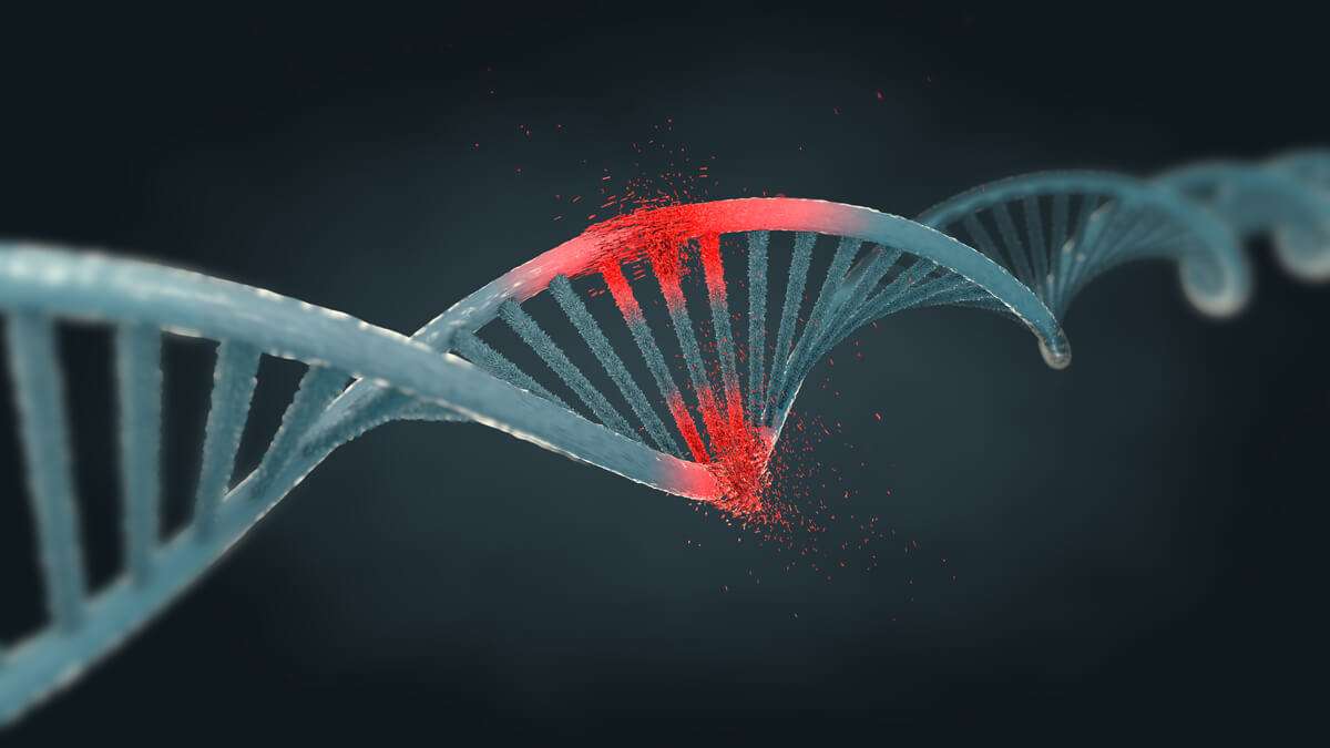
Cells constantly deal with damage to their DNA that can originate from endogenous and exogenous sources, such as replication stress, telomere shortening, UV radiation or chemical toxins. DNA damaging agents give rise to different types of DNA damage, and failure to repair the damage can result in several possible devastating consequences to the cell, including mutations, genomic instability and even cell death contributing to many diseases, including cancer.
Cells have therefore developed intricate repair mechanisms, collectively referred to as the DDR to deal with the different types of possible DNA lesions that arise; all with the goal of protecting genomic stability.

The major types of DNA repair mechanisms are:
Direct Reversal
Direct reversal represents the simplest form of DNA repair since it depends mostly on the activity of a single protein without the need for nucleotide removal, resynthesis, or ligation. For instance, O6-methylguanine (O6-mG) is a harmful genetic lesion induced by alkylating mutagens. The presence of alky groups at the O6 position of guanine causes G:C to A:T transitions and a block in gene transcription or DNA replication. In the direct reversal process, the alkyl group is removed by the enzyme O6-methylguanine DNA methyltransferase (MGMT) and the proper nucleotide is restored.
Base Excision Repair (BER)
BER is a highly conserved mechanism, spanning from bacteria to human, responsible for repairing small but highly mutagenic DNA lesions that are a significant threat to genomic fidelity and stability. These DNA lesions can be caused by ionizing radiation as well as endogenous mutagens generated from metabolic events such as oxidation, methylation, deamination, or spontaneous loss of a DNA base pair. The BER pathway is initiated by one of eleven DNA glycosylases, each with specificity for distinct types of DNA damage. These enzymes remove the damaged base resulting in abasic (apurinic/apyrimidinic) sites, also known as AP sites. Following base excision, a repair process involving DNA polymerases and DNA ligases will play a crucial role, filling the exposed gap with a single base (short-patch pathway) or multiple bases (long-patch pathway).
Nucleotide Excision Repair (NER)
NER is perhaps the most flexible mechanism in terms of the diversity of lesions that are recognized and repaired. The most significant of these lesions are pyrimidine dimers induced by UV light and other NER substrates include bulky chemical adducts, DNA intra-strand crosslinks, and some forms of oxidative damage. These types of lesions cause both a helical distortion of the DNA duplex and a modification of the DNA chemistry, both of which are hallmark features recognized by NER.
NER is a complex multi-step process involving a large network of more than 30 proteins. Initiated by recognition of the damaged base, the DNA duplex is then unwound, and the damaged base(s) is removed by an excision repair complex which is then filled in and ligated.
DNA Mismatch Repair (MMR)
MMR is also a very highly conserved biological pathway, whose main function is to eliminate base pair mismatches and minor insertions/deletions that occur due to replication errors and spontaneous or induced alterations in bases. MMR also corrects mismatched pairs or “mispairs” generated by the spontaneous deamination of 5-methylcytosine and heteroduplexes formed following genetic recombination.
Defects in this pathway result in a "mutator" cellular phenotype characterized by elevated frequencies in spontaneous mutations and increased microsatellite instability (MSI). Mutations in several human MMR genes cause a predisposition to hereditary nonpolyposis colorectal carcinoma (HNPCC), resistance to certain chemotherapy drugs as well as a variety of sporadic tumors that display MSI.
Double-Strand Break (DSB) Repair
DSBs are highly toxic genetic lesions that pose a serious threat to cellular homeostasis since they can affect transcription, replication, and chromosome segregation. DSBs are caused by a variety of exogenous and endogenous factors, such as ionizing radiation, certain genotoxic chemicals, and reactive oxygen species.
DSBs differ from most other types of DNA lesions mostly because they affect both strands of the DNA and therefore prevent use of the complementary strand as a template for repair. Failure to repair DSBs can result in devastating chromosomal instabilities that can lead to dysregulated gene expression, chromosomal translocations and increased risk of carcinogenesis.
Cells have evolved into 4 distinct pathways of DSB repair: homologous recombination (HR), non-homologous end joining (NHEJ), alternative end-joining (alt-EJ) and single-strand annealing (SSA) but HR and NHEJ are the two most important. While a cell may opt to use either of these pathways to repair DSBs, the exact details of why a cell chooses one pathway over another remains unknown. Selection appears to be influenced by the cell cycle stage at the time the damage was acquired.
Homologous Recombination (HR)
HR maintains genome stability by repairing DSBs, gaps, and restarting stalled replication forks. It is a relatively slow but error-free pathway that relies on the presence of a homologous sequence in the genome as a template to replace the damaged DNA segment. It primarily occurs during the S and G2 phases of the cell cycle. HR is a relatively complex process involving a large number of proteins, including the PI3K kinase family member ataxia-telangiectasia mutated (ATM). ATM is a critical regulator in several signaling cascades. Upon DNA damage, it becomes monomeric, phosphorylating itself on different Serine. Mutations in the phosphorylation sites can result in defects in the ATM signaling pathway, leading to radiation sensitivity for instance.
Non-Homologous End Joining (NHEJ)
Unlike HR, NHEJ does not require a homologous DNA template for repairing DSBs. Instead, NHEJ operates by modifying the free ends of DNA located on either side of the break by using various nucleases so that the ends become compatible followed by ligation with the enzyme DNA ligase 4. In contrast to HR, NHEJ is a relatively quick but intrinsically error-prone process, and its excessive use can lead to gene rearrangements, deletions, and mutations, all of which can cause post-replicative cells to be more vulnerable to DSBs.
DNA damage sensor proteins and DNA damage signaling proteins can both be targeted with a range of anticancer agents.
Drug Targeting of DNA Damage Sensor Proteins
DDR is essential for the activation of repair pathways and cell survival. DDR sensor proteins, which respond to a wide variety of DNA lesions, are key for initiating repair. For DSBs, Ku and MRN represent the major sensor protein complexes.
Ku is a protein heterodimer consisting of Ku70/Ku80, and it is part of the NHEJ machinery that immediately binds to DNA DSBs. Upon recognition and binding of DSBs, Ku recruits other proteins that assist in classical NHEJ repair.
The MRN complex is also important for initial detection of DSBs. After binding to the damaged site, MRN recruits the DNA-damage signaling kinase ATM where it becomes activated and triggers a cascade of signaling events that initiate DNA end-resection and promote repair by HR. A variety of other DNA-damage sensors have also been identified including the Fanconi anemia core complex, mismatch repair proteins, nucleotide excision repair proteins which are sensors of DNA inter-strand crosslinks, base-base mismatches or insertion-deletion loops and UV-induced photo-lesions.
PARP-1 is a key DNA damage sensor protein that has been successfully targeted with drugs largely due to the synthetic lethal relationship between PARP and BRCA. Synthetic lethality arises when simultaneous loss of function of two genes (often in the same pathway) results in cell death. The clinical success of targeting of PARP with small molecule inhibitors (e.g. olaparib, talazoparib, niraparib, rucaparib, and velaparib) in BRCA-mutated ovarian cancer has provided proof-of-principle for exploiting synthetic lethality as a treatment strategy and has led to the development of other therapies targeting the DNA damage response.
Drug Targeting of DNA Damage Signaling Proteins
DDR signaling proteins trigger a wide variety of post-translational modifications and the assembly of protein complexes that amplify and diversify the DNA damage signal so that the appropriate responses can be initiated, and can include: transcriptional changes, cell cycle checkpoint activation, alternative splicing, engagement of DNA repair processes, or in the context of massive damage, activation of cell senescence or apoptotic pathways.
The main proteins coordinating the signaling events of the DDR and novel drugs targeting this pathway are discussed below.
ATM/ATR
ATM is a protein kinase that facilitates DSB repair and responds to DSBs throughout the cell cycle. ATM is predominantly activated through interactions with NBS1 of the MRN complex. It is the principal kinase responsible for the phosphorylation of histone H2AX, which occurs rapidly following DSBs and serves as a foundation for the assembly of the DNA repair machinery. ATM inhibition has been demonstrated to make cells very sensitive to ionizing radiation and DSB-inducing agents (e.g., etoposide, camptothecin, and doxorubicin). Ongoing research and development have led to the investigation of numerous ATM inhibitors in the field of oncology. A notable example is AZD0156, which is currently under a phase I clinical trial. This trial is evaluating its efficacy as a standalone treatment and in a synergistic application with the PARP inhibitor olaparib, alongside other cytotoxic agents such as irinotecan.
ATR is activated by replication protein A (RPA) bound at sites of ssDNA such as stalled replication forks or following 5’-3’degradation of one of the DNA strands (i.e., DNA-end resection) during the early stages of HR. In recent developments, a range of ATR inhibitors have progressed to clinical trials, including M6620 (also known as VX-970 or berzosertib), M4344 (VX-803), AZD6738, and BAY1895344. Of particular interest is the ATR inhibitor, M6620, which has demonstrated potential both as a single agent and when used in combination with carboplatin. Furthermore, NU6027 has emerged as a significant ATR inhibitor, amplifying the cytotoxic effects of hydroxyurea and cisplatin and functioning through an ATR-dependent mechanism.
DNA-PK
DNA protein kinase (DNA-PK) is required for proper DSB repair by NHEJ, which is the primary DNA repair pathway for DSBs in human cells that occurs through all phases of the cell cycle. Small molecule inhibitors of DNA-PK kinase activity stabilize DNA-PK on DNA ends and subsequently impair NHEJ, likely also interfering with other repair processes, including HR, by obstructing DNA end-resection. Loss of DNA-PK activity leads to reduced cell proliferation and the initiation of caspase-mediated cell death. One of the challenges associated with targeting DNA-PK has been selectivity due to structural similarities with other kinases. Given its role in NHEJ, drugs that target DNA-PK have been found to be more effective when combined with agents that induce replication-independent DSBs, such as ionizing radiation and topoisomerase 2 inhibitors (e.g., doxorubicin, etoposide).
A number of DNA-PK inhibitors are available, including NU7441, nedisertib, AZD7648, VX-984, berzosertib, ceralasertib, VX-803, BAY1895344, CC-115, NU7427, NU7026, and NU7163. AZD7648 is in the early stages of clinical testing (phase I) and has been shown to sensitize cells to radiation- and doxorubicin- induced DNA damage. Simultaneously, VX-984 is undergoing phase I clinical trials and has been found to improve the response of glioblastoma cells to radiation therapy. In the realm of leukemia treatment, NU7026 has demonstrated an ability to enhance the effectiveness of drugs that target topoisomerase II poisons.
CHK1/2
CHK1 is a protein kinase that lies downstream of ATR and functions as a critical regulator of the intra-S and G2-M cell cycle checkpoints. Given the role of CHK1 in mediating cell cycle arrest following DNA damage, it is not surprising that CHK1 inhibitors appear to be most effective when combined with agents inducing DNA damage during DNA replication.
Among the CHK1/2 inhibitors being investigated, Rabusertib (LY2603618) has emerged as a good candidate. It is a highly selective, small molecule inhibitor targeting the CHK1 protein kinase, and it was among the earliest of such inhibitors to enter clinical cancer trials. LY2603618, when administered in combination with pemetrexed and cisplatin, demonstrated an acceptable safety profile. MK-8776 (previously known as SCH 900776) has shown potential both as a standalone chemotherapeutic agent, and when used in combination with DNA antimetabolites such as pemetrexed and gemcitabine.
WEE1
The WEE1 protein kinase, which works in parallel with CHK1, plays a critical role in activating the G2-M checkpoint through the regulation of cyclin dependent kinases. However, unlike CHK1, WEE1 is not directly regulated by DNA damage but it is required for physiological cell cycle progression. As for WEE1 inhibitors, it was believed that their mechanism-of-action led to mitotic catastrophe by preventing the activation of the G2-M checkpoint as a result of inappropriate CDK1/CCNB1 activation. However, more recent data have demonstrated that WEE1 inhibition also generates replication-dependent DNA damage in cells due to aberrant DNA replication through CDK2 inhibition.
The WEE1 protein kinase, which works in parallel with CHK1, plays a critical role in activating the G2-M checkpoint through the regulation of cyclin dependent kinases. However, unlike CHK1, WEE1 is not directly regulated by DNA damage but it is required for physiological cell cycle progression. As for WEE1 inhibitors, it was believed that their mechanism-of-action led to mitotic catastrophe by preventing the activation of the G2-M checkpoint as a result of inappropriate CDK1/CCNB1 activation. However, more recent data have demonstrated that WEE1 inhibition also generates replication-dependent DNA damage in cells due to aberrant DNA replication through CDK2 inhibition.
DNA Pol β
DNA Pol β is a key player in both nuclear and mitochondrial BER pathways. Pol β also plays a part in repairing DSBs via the alt-EJ pathway. In the 1990s, Mizushina and his team found that linoleic acid (LA), a commonly known fatty acid, was capable of inhibiting calf thymus DNA pol α and rat DNA pol β. Following this discovery, over the next twenty years, Mizushina and other researchers identified a wide array of Pol β inhibitors. These included a range of compounds, including polypeptides, fatty acids, triterpenoids, sulfolipids, polar lipids, phenalenone derivatives, flavonoid derivatives, etc. However, the effectiveness and specificity of most of these inhibitors have been too limited for them to be developed into approved medicinal drugs. Consequently, future research should explore a broader approach for the discovery of novel Pol β inhibitors that not only offer reduced side effects but also have better potential for use as therapeutic agents.
ERCC1
The NER pathway is integral in removing large DNA adducts that are a result of factors like UV radiation, external agents, lipid peroxidation, or reactive oxygen species (ROS). These adducts lead to a distortion of the DNA’s helical configuration, which can trigger cell cycle arrest and induce cell death via apoptosis. Clinically, drugs such as platinum-based cisplatin use this pathway to target cancer cells. However, in many cancer cells, the NER pathway is overactive due to the overexpression of ERCC1, which can induce drug resistance and therefore diminish the therapeutic effectiveness of cisplatin. Consequently, crafting inhibitors that selectively target NER/ERCC1 holds promise for the enhancement of the efficacy of platinum-based treatments.
WRN
The Werner syndrome ATP-dependent helicase (WRN), part of the RECQ family of DNA helicases, plays a crucial role in the unwinding of double-stranded DNA for both DNA replication and repair processes. WRN uniquely possesses 30 to 50 exonuclease activities in addition to its helicase activity. Individuals who inherit germline mutations in the WRN gene present characteristics of Werner syndrome, such as increased genomic instability and a higher risk of developing cancer.
DNA Damage Response Combination Strategies
The ability to effectively respond to DNA damage is often lost in many types of cancers and targeting this pathway has been clinically validated in subsets of patients, such as those with BRCA1/BRCA2 mutations treated with a PARP inhibitor. To enhance therapeutic efficacy, DDR inhibitors can be combined with drugs targeting other DDR proteins or completely different signaling pathways, with the goal of blocking multiple pathways that cancer cells rely on for survival.
For example, PARP inhibitors trap PARP1 and 2 on DNA strands, generating DNA adducts and stalled replication forks which require ATR for resolution. Therefore, combined PARP and ATR inhibition holds potential to further increase replication stress and induce cell death. Yet advancing combination therapies in practice presents its own set of complexities. Initial studies, such as those in a small phase II trial for ovarian cancer, show promise but highlight a pressing need for refinement. One of the primary challenges is managing the overlapping toxicities of combined ATR and PARP inhibitors, requiring development strategies that balance tolerability while preserving treatment efficacy. Innovations such as sequential dosing of DDR inhibitors and more precisely targeted DDR inhibitors might be key advancements for combined therapeutic strategies.
Combination therapy research has expanded to incorporate different chemotherapies, including platinum salts, gemcitabine, topoisomerase inhibitors, and taxanes, which have been evaluated with ATR or WEE1 inhibitors in preclinical and clinical settings. Specifically, combination therapy using non-platinum chemotherapy classes with ATR/WEE1 inhibitors has shown higher tolerability. Promising results have been noted with particular combinations—gemcitabine with adavosertib or berzosertib, paclitaxel with ceralasertib, and topotecan plus berzosertib.
Combining DDR inhibitors with antibody-drug conjugates (ADCs) has surfaced as an attractive strategy since these combinations tend to have fewer overlapping toxicities. For example, pyrrolobenzodiazepine dimers (PBD) are alkylators that form intra-strand cross-links within the DNA minor groove, leading to dsDNA breaks, and are a potent ADC payload. A pertinent and ongoing trial is looking at the novel PARP1-selective inhibitor, AZD5305, in combination with the human epidermal growth factor receptor 2 (HER2)-targeting ADC, trastuzumab deruxtecan, or the Trop2-targeting ADC, datopotamab deruxtecanin, in advanced cancer patients with selected HRR gene mutations. The potential for other DDR inhibitor and ADC combinations is also being explored.
Emerging scientific and clinical evidence is indicating a potential synergy between the body’s DDR and immune responses. Leveraging knowledge of the interactions between DNA damage, DDR, and the immune response could lead to an increase in the clinical efficacy of immunotherapies by combining them with DDR inhibitors and/or radiation as sensitizers. In particular, combinations that lack overlapping drug-related toxicities can be advantageous, potentially enabling full monotherapy doses of both drugs to be administered concurrently. Current research is probing combinations like ATR, WEE1, and other DDR inhibitors with immune checkpoint blockade inhibitors.
Resistance to DNA Repair Inhibitors in Cancer
Insights gained from the development and resistance studies of PARP inhibitors (PARPis) and the study of PARPi resistance could inform how to circumvent resistance in the clinical development of new DDR inhibitors. Several mechanisms by which cancers develop resistance to PARPis have been identified, including reversion mutations in genes like BRCA1/2, PALB2, RAD51C, and RAD51D, which restore the gene’s reading frame, allowing the encoding of functional proteins. Even though reversion mutations have been identified in a substantial number of patients resistant to PARP inhibitors, a significant segment of the PARPi-resistant patient community still has unidentified reasons for their resistance to the treatment.
Further pre-clinical studies indicate that alterations in a series of other DNA repair proteins cause PARPi resistance by restoring HR without the necessity for genetic reversion of a mutant BRCA1/2 allele (53BP1, RIF1, SHLD1, SHLD2, SHLD3, REV7, the CST complex, PTIP, EZH2, DYNLL1, SLFN11, etc.) or changes in the drug target.
With ATR inhibitors (ATRis) being a more recent entry into clinical development, insights into ATRi resistance primarily come from pre-clinical genetic perturbation screens. These studies suggest that ATRi resistance might occur due to the loss or mutation of certain proteins, like Cyclin E, CDK2, or Myc, or through mechanisms that induce cell stalling, such as the loss of CDC25A/B phosphatase activity or the loss of the CDK8/Cyclin C complex. Similarly, mutation of the pro-mitotic transcription factor FOXM1 has been linked to ATRi resistance.
In general terms, this means that particular attention must be paid to the main mechanisms, such as mutations in the drug target itself, pharmacokinetic (PK) resistant mechanisms, or changes in pathways that compensate for the loss of activity, to avoid the development of resistance mechanisms as much as possible.
Conclusion and perspectives
While cancer cells may benefit from defects in the DDR pathway, the presence of other functioning DNA repair systems can enhance survival. Drugs that target the DDR have been pre-clinically and clinically validated in subsets of patients, and different combination strategies are actively under study to inhibit multiple pathways that cancer cells rely on for survival.
Leveraging PDX models with a high level of characterization holds promise for the future discovery of new combinations that offer efficacy with minimal side effects, alongside the identification of new biomarkers. Illustratively, a study employing a panel of 11 PDX models demonstrated a great activity of DDRi agents used in combination, specifically within models with biallelic ATM mutations and associated protein loss.

By impeding DNA repair, DDR inhibitors are ideally suited to combination therapies that can improve the efficacy of radiotherapy, chemotherapy, or immunotherapy.
Further Reading
Yi et al. Advances and perspectives of PARP inhibitors. Experimental Hematology and Oncology 2019;8: 1–12.
Helleday et al. DNA repair pathways as targets for cancer therapy. Nature Reviews Cancer 2008;8:193–204.
Brown et al. Targeting DNA repair in cancer: Beyond PARP inhibitors. Cancer Discovery 2017;7: 20–37.
Mouw et al. DNA damage and repair biomarkers of immunotherapy response. Cancer Discovery 2017;7:675-93.
Ngoi° et al. Targeting the DDR beyond PARP inhibitors: Novel agents and rational combinations. Curr Opin Oncol. 2022.
Zou et al. DNA repair defects in cancer and therapeutic opportunities. Genes and Development 36:278–293.
Baxter et al. Resistance to DNA repair inhibitors in cancer. Molecular Oncology 16 (2022) 3811–382.
Concannon et al. Combining Targeted DNA Repair Inhibition and Immune-Oncology Approaches for Enhanced Tumor Control Mol Cell. 2023 March 02; 83(5): 660–680.
Wang et al. Targeting the DNA Damage Response for Cancer Therapy Int. J. Mol. Sci. 2023, 24, 15907.