Podcast
The Power and Potential of Biomarkers
Oncology continues to be a rapidly changing field with scientific discoveries transforming patient treatment and care. As it stands today, advancement…

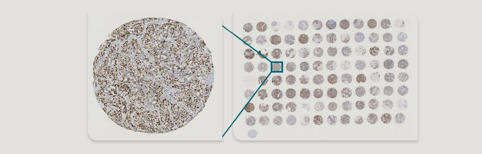
Advancements in oncology are increasingly driven by the ability to extract more detailed and precise information from patient tumor samples. Traditional histopathological techniques, while foundational, often fail to capture the spatial heterogeneity and cellular interactions that underlie complex tumor microenvironments. Enter spatial data analysis—a transformative approach that brings context back into molecular analysis by preserving the geographic architecture of cells within tissue samples.
As researchers strive to uncover novel biomarkers and develop targeted therapies, the integration of spatial data analysis with patient tumor samples has emerged as a new frontier. This convergence is not just enhancing our understanding of cancer biology but is also redefining biomarker discovery and translational medicine.
Spatial data analysis enables a multidimensional perspective that reveals how the spatial arrangement of cells and molecular features influences tumor progression, immune infiltration, and therapeutic response. Instead of relying solely on bulk molecular signals, which average out critical differences between regions of a tumor, spatial technologies empower researchers to zoom in on specific cellular neighborhoods and study their functional roles. This spatially informed perspective is essential for unraveling the complexity of tumor ecosystems, which often consist of diverse cell populations with varying degrees of malignancy, immune activity, and treatment sensitivity.
The implications for personalized medicine are profound. By characterizing the spatial dynamics of tumor-immune interactions, vascularization, and metabolic gradients, clinicians can develop more precise diagnostic tools and tailor treatment strategies to each patient’s unique tumor architecture. Spatial data analysis holds the promise of turning traditional biopsy samples into rich, layered maps that guide oncologists in selecting the right therapies at the right time—ultimately improving outcomes and reducing unnecessary interventions.
Spatial data analysis refers to the computational and statistical examination of spatially resolved biological data. Unlike bulk sequencing, which homogenizes a tissue sample, or single-cell RNA sequencing, which dissociates cells from their spatial niches, spatial technologies maintain the locational context of each cell within a tissue section. This enables researchers to map gene expression, protein localization, and cell-cell interactions with spatial precision.
When applied to patient tumor samples, spatial data analysis provides a multidimensional view of the tumor microenvironment (TME), capturing complex spatial arrangements and revealing insights that would otherwise remain hidden.
By retaining spatial information, researchers can dissect how distinct cell populations are organized and how their proximity—or lack thereof—impacts biological function. For example, spatial analysis can show whether immune cells are actively infiltrating a tumor or are confined to its periphery, an important distinction that can influence the effectiveness of immunotherapies. It can also reveal patterns of cell signaling or metabolic activity localized to specific regions of a tumor, allowing scientists to study the microenvironmental niches that drive tumor aggression or resistance.
Additionally, spatial data analysis facilitates a better understanding of tumor architecture over time and space. It supports longitudinal studies where researchers can compare spatial features in pre- and post-treatment samples to identify biomarkers associated with response or resistance. This capability is critical in a clinical setting, where identifying actionable spatial biomarkers could inform therapeutic decisions and provide a dynamic view of how tumors evolve under therapeutic pressure.
Several cutting-edge platforms are propelling this field forward:
Together, these technologies generate spatially resolved datasets that are large, complex, and information-rich—perfect for advanced analytics and integrative biomarker discovery.
The integration of these tools has also led to the rise of spatial multi-omics, where transcriptomics, proteomics, and metabolomics data can be captured from the same tissue sample, sometimes even at single-cell resolution. This convergence allows researchers to link gene expression to protein localization and cellular function within the precise architecture of the tumor. As a result, spatial multi-omics platforms are unlocking new avenues for biomarker discovery and therapeutic target validation.
Moreover, advancements in tissue processing now allow spatial technologies to be applied to formalin-fixed paraffin-embedded (FFPE) samples—the most commonly archived tissue type in clinical settings. This compatibility means researchers can conduct retrospective spatial analyses on large biobanks of patient tumor samples, facilitating longitudinal studies, cross-cohort comparisons, and clinical trial support. These innovations are bringing spatial data analysis out of the research lab and into the realm of real-world, translational oncology.
The TME comprises malignant cells, immune infiltrates, fibroblasts, vasculature, and extracellular matrix components—all dynamically interacting in a spatially organized manner. Spatial data analysis enables:
By visualizing these interactions within their native spatial context, researchers can assess how the positioning of various cell types influences tumor behavior. For instance, the proximity of cytotoxic T cells to cancer cells may indicate an active immune response, whereas a physical barrier of fibroblasts or an immunosuppressive milieu could suggest immune exclusion. This level of detail provides a foundation for designing therapies that not only target tumor cells but also modulate the supporting environment to enhance treatment efficacy.
Patient tumor samples often exhibit substantial intratumoral heterogeneity—variations in cell types, genetic mutations, and microenvironmental conditions. Spatial data analysis uncovers:
These spatial variations are often predictive of treatment response or resistance. For example, hypoxic regions within a tumor may harbor cells that are more resistant to chemotherapy or radiotherapy. Similarly, spatial patterns of stromal or immune cell infiltration can guide the use of immunotherapies. By visualizing these features, researchers gain a nuanced understanding of tumor biology that is critical for therapeutic development.
Furthermore, spatial analysis enables the identification of evolutionary dynamics within tumors, such as the expansion of resistant clones or the emergence of new phenotypes under therapeutic pressure. This has significant implications for real-time monitoring of disease progression and adaptive treatment strategies, ensuring that interventions remain aligned with the tumor’s evolving landscape. Ultimately, integrating spatial data into cancer research is accelerating the shift toward precision oncology, where therapy is informed not only by molecular signatures but also by spatial tissue architecture.
Traditional biomarker discovery often relies on measuring the average gene or protein expression across an entire tissue sample or patient cohort. While this bulk approach has yielded valuable insights, it overlooks critical spatial heterogeneity—where the location of specific molecules and cells within the tissue profoundly influences function and therapeutic outcomes. Spatial data analysis disrupts this paradigm by providing context-aware biomarker profiles, focusing on where molecular features occur and how they interact with their surrounding environment.
Spatial patterns, such as immune cell exclusion or the presence of tertiary lymphoid structures (TLS), have been correlated with patient survival across several cancer types. TLS, often found near tumor margins, are associated with improved prognosis in certain solid tumors due to their role in orchestrating local immune responses. These complex spatial configurations are invisible to bulk profiling but are clearly identified through spatial transcriptomics and multiplexed imaging, making them powerful prognostic indicators.
Checkpoint blockade therapies (e.g., anti-PD-1/PD-L1) have revolutionized cancer treatment, particularly in melanoma, non-small cell lung cancer, and renal cell carcinoma. However, many patients fail to respond. Spatial data from patient tumor samples has revealed that the proximity between T cells and tumor cells—not merely their presence—serves as a stronger predictor of therapeutic response. For example, an inflamed tumor phenotype with T cells infiltrating the tumor core is more likely to respond favorably, compared to tumors where immune cells are restricted to the periphery.
In drug development, spatial analysis facilitates stratification of responders versus non-responders by identifying microenvironmental features such as stromal barriers, vasculature proximity, and immune cell clustering. These structural insights help optimize trial cohorts, guide dosing strategies, and enable more efficient development of companion diagnostics. Furthermore, spatially resolved markers can inform mechanisms of drug resistance, such as the emergence of immune escape niches or the remodeling of tumor architecture post-treatment.
Incorporating spatial biomarkers into clinical trials also allows for real-time tissue monitoring. Biopsy samples collected at different time points can be spatially profiled to assess how tumors evolve in response to therapy, providing critical feedback for treatment adjustments. As spatial technologies become more standardized and scalable, they are expected to play an increasingly central role in precision oncology workflows, guiding both therapeutic development and clinical decision-making.
Spatial data analysis doesn’t exist in isolation—it thrives when integrated with other layers of biological and clinical information to provide a comprehensive understanding of cancer biology. This synergistic approach is essential for unraveling the complexity of tumor behavior and enhancing translational relevance.
In the future, integrating spatial data with longitudinal clinical records and liquid biopsy results could support dynamic tumor monitoring—offering a continuous feedback loop to guide treatment decisions. Multi-modal spatial atlases, enriched with clinical annotations, are already being used to power machine learning models that predict outcomes and therapeutic response. This convergence of spatial analysis, multi-omics, and clinical metadata represents a critical step toward realizing the full potential of precision oncology.
While promising, spatial data analysis in oncology faces several hurdles that must be addressed to fully realize its potential in research and clinical practice:
Nonetheless, the field is advancing rapidly. Emerging trends include:
Looking ahead, democratizing access to spatial technologies through cost reduction, user-friendly software, and cloud-based analytics will be key to accelerating adoption in both academic and clinical settings. Furthermore, collaborative initiatives such as the Human Tumor Atlas Network (HTAN) and Cancer Moonshot are creating publicly available spatial atlases and benchmarking datasets that will help standardize the field and accelerate discoveries. Spatial data analysis is poised not only to redefine how we study cancer but also to transform how we diagnose, monitor, and treat it.
The integration of spatial data analysis with patient tumor samples marks a transformative step in cancer research and clinical practice. By preserving the spatial architecture of tumors, researchers can unlock new insights into tumor biology, uncover spatial biomarkers, and accelerate the development of personalized therapies. This approach moves beyond isolated molecular profiles to deliver a comprehensive view of how cells interact, adapt, and evolve within their native microenvironments.
This frontier promises not just deeper scientific understanding but also tangible improvements in patient outcomes. Spatial analysis enables the development of more effective diagnostic tools, enhances patient stratification in clinical trials, and informs targeted treatment strategies that are tailored to the spatial and molecular complexity of individual tumors. The ability to map therapeutic vulnerabilities within distinct tumor niches paves the way for more precise interventions with fewer off-target effects.
As spatial technologies become more accessible, automated, and integrated with digital pathology and electronic health records, their utility in routine clinical workflows will grow. We can envision a future where spatially guided diagnostics inform real-time treatment decisions, spatial biomarkers are standard in pathology reports, and multi-omic spatial maps serve as a digital fingerprint for every tumor. In this way, spatial data analysis is not just a technological advancement—it is a paradigm shift that is reshaping the landscape of oncology, bringing us closer to truly personalized cancer care.

Crown Bioscience, (2025) - Crown Bioscience. https://blog.crownbio.com/spatial-data-analysis-patient-tumor-samples