Explore the utility of preclinical mouse models of metastasis to study circulating tumor cells (CTCs) and the techniques used to isolate and characterize them.
We’ve previously posted on which preclinical mouse models best recapitulate human metastatic disease progression. In this post, we’re focusing on the use of these models to study circulating tumor cells, including the latest techniques to isolate and characterize CTCs. Isolation and characterization of CTCs at the molecular and functional level shows great promise for therapeutic development in oncology.
Modelling Circulating Tumor Cells In Vivo
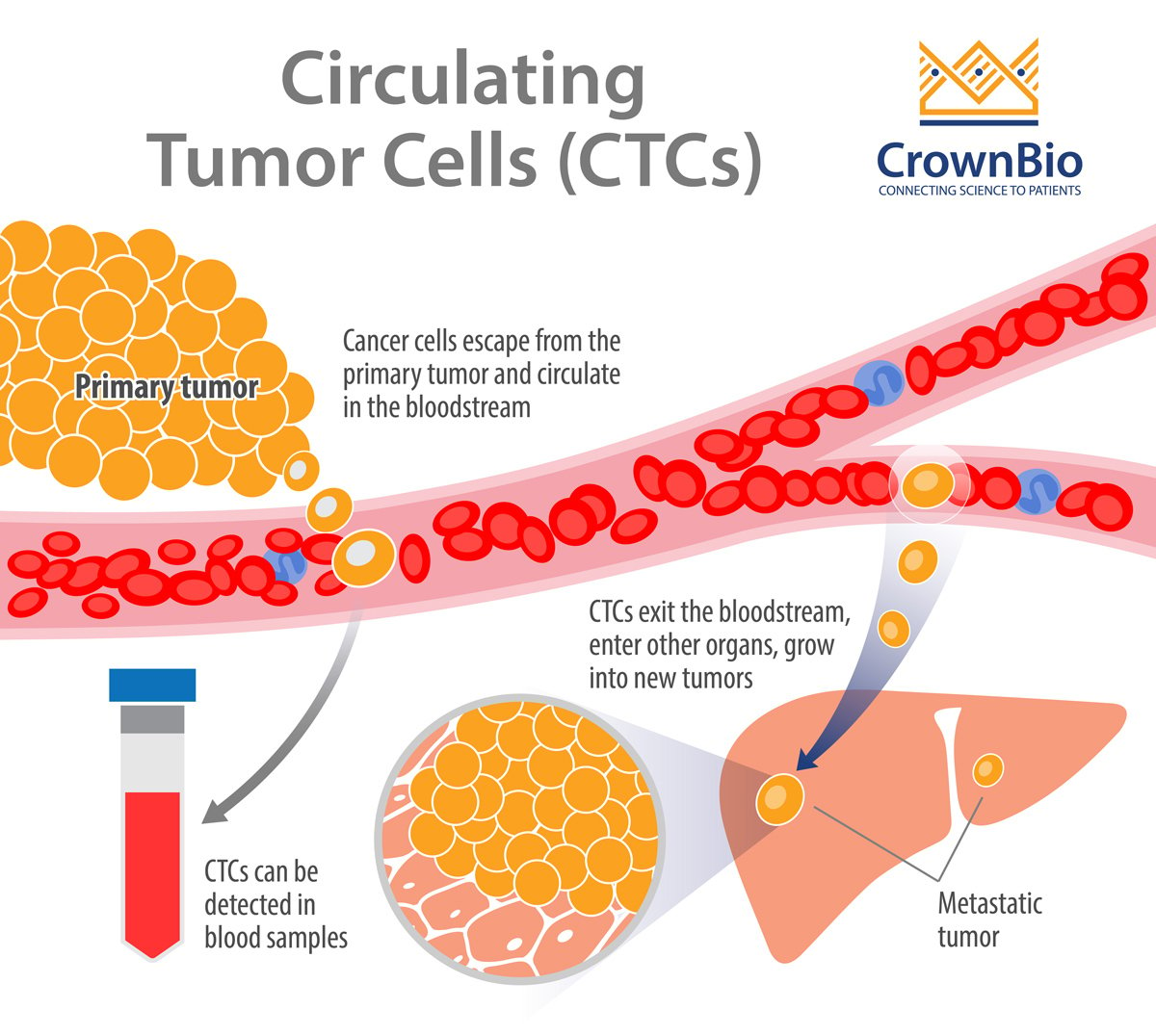
At a very basic level, CTCs are cancer cells that have been shed from a primary tumor or metastatic site, which circulate through blood and/or lymphatic vessels as single cells or aggregates. A subset of CTCs survives, extravasates into the interstitial space, and finally forms a tumor in a new and distant microenvironment.
The term “liquid biopsy” was coined to describe the process of detecting, characterizing, and monitoring non-hematological cancers based on CTCs isolated from peripheral blood. Liquid biopsy has attracted a lot of attention from a variety of stakeholders, including drug developers and clinicians. This is because CTCs can serve as drug targets, guide therapeutic cancer care (e.g. evaluate treatment efficacy, adjust treatment based on molecular profile, monitor for residual disease), and reduce the burden associated with traditional tissue biopsies.
Preclinical Models of CTCs
To study CTCs in preclinical mouse models, metastatic disease must be generated in vivo. There are a variety of metastatic models available, each with advantages and disadvantages when it comes to best recapitulating the progression of the human metastatic process.
Once the ideal metastasis model is selected, CTC analysis provides a unique opportunity for developing new biomarkers and characterizing and tracking metastatic disease progression, which can provide key insights for novel clinical treatments. For these reasons, preclinical metastasis models are increasingly being used to study CTCs and are also being directly established using CTCs isolated from patient blood samples or after ex vivo expansion.
Breast Cancer
CTCs were identified in mice using patient-derived xenograft (PDX) models of breast cancer. This type of model provides a constant and renewable source of CTCs, allowing for the study of CTCs and underlying metastatic processes.
CTC cell lines have also been established from breast cancer CTCs and used to identify a potential signature of brain metastasis. CTCs with this signature were found to be highly invasive and generated brain and lung metastases when xenografted into immunocompromised mice. Similarly, breast cancer CTCs were isolated from patients with estrogen receptor–positive breast cancer and used to establish cell lines, 60% of which were tumorigenic in mice.
Lung Cancer
Patient-derived CTCs are being used to generate preclinical mouse models with CTC-derived explants, including for small-cell lung cancer (SCLC), which is known to harbor high levels of CTCs. For instance, patient-derived SCLC CTCs were tumorigenic in immunocompromised animals, and the CTC-derived model mirrored donor patient response to chemotherapy.
Colon Cancer
A permanent cell line from CTCs derived from a colon cancer patient was established sharing the characteristics of the original tumor, and which induced tumors in immunocompromised animals allowing functional studies and drug evaluation.
Enrichment and Characterization of CTCs
Before a CTC study, researchers should familiarize themselves with the techniques commonly used to enrich and characterize CTCs. In particular, enrichment remains a major technical challenge because CTCs are:
Rare in the peripheral blood (estimated to occur at ~1 CTC per 105–108 white blood cells).
Suspected to have a short half-life.
Accompanied by a paucity of reliable surface biomarkers.
However, recent technological advances have helped circumvent some of the challenges and there are commercially available platforms that collect and identify CTCs.
CTCs are usually identified as nucleated cells that express epithelial markers cytokeratin (CK)/EpCAM but not CD45. However, the current drawback of capturing CTCs by the recognition of epithelial-specific markers is the lack of ability to detect CTCs that undergo epithelial-mesenchymal transition (EMT) and no longer overexpress EpCAM. CTCs undergoing EMT acquire mesenchymal morphology which is associated with the apparition of the expression of mesenchymal markers such as vimentin (CSV), Slug, or Twist, that can be use as alternative or complementary methods able to recognize a broader spectrum of phenotypes.
CTC Capture
Immunomagnetic-based enrichment is the most commonly used technique to capture CTCs. It relies on magnetic, bead-based separation technology, where CTCs are enriched from blood samples using antibodies conjugated to magnetic beads. The antibodies select for CTCs either positively, by targeting epithelial or tumor-specific antigens, or negatively by targeting blood cell antigens (e.g. CD45).
Multiple commercial vendors market different products. Notably, some of the positive selection products, such as the EpCAM based systems, target only epithelial cells. Today the CellSearch® CTC Kit is the only FDA-approved strategy to detect CTC of epithelial origin (CD45-, EpCAM+, and cytokeratins 8, 18+, and/or 19+) in whole blood.
Density-based enrichment relies on the CTCs being generally less dense than blood cells. There are several commercial vendors of density gradient media that separate CTCs from blood cells. This technique captures both epithelial and mesenchymal CTCs and is generally simple to use and inexpensive, however the method shows low specificity.
Size-based enrichment relies on the difference in size between CTCs and white blood cells. CTCs are larger than leukocytes and filtration devices have pore sizes that separate the two. This technique captures both epithelial and mesenchymal CTCs and is also generally simple to use and inexpensive. Nevertheless, CTCs of smaller sizes may be lost.
Microfluidic-based enrichment involves passing whole blood through microchannels on chip-based devices. As CTCs travel through the channels, they are captured by antibodies attached to “microposts” or filtered by size. Multiple microfluidic options are commercially available. These devices can handle fluid volume in the µL to nanoliter range, and they generally allow for high enrichment and isolation of intact CTCs.
CTC Characterization
Regardless of the enrichment technique, subsequent downstream characterization (i.e. detection, enumeration, and/or molecular characterization) is required, which is possible using a variety of protein- and/or nucleic-acid-based approaches.
Protein-based detection and molecular characterization is performed via one or more of several techniques. Common options include flow cytometry and slide-based immunofluorescence, which rely on fluorescent-conjugated antibodies that target antigens on CTCs. These are then identified with standard laser- or image-based detection systems.
Nucleic acid-based detection and molecular characterization is typically performed with real-time and quantitative real-time PCR, which identify CTCs based on expression of specific genes. Other techniques, such as next-generation sequencing and other genomic analyses, are increasingly being used. Notably, these assays don’t allow for accurate quantification of CTCs or recovery of CTCs for additional analysis.
In addition to CTCs, liquid biopsies also offer the opportunity to evaluate ctDNA. That is another circulating biomarker that has high sensitivity and may be useful in treatment selection, response monitoring, and monitoring tumor burden and molecular features of disease. Isolation and analysis of ctDNA can be technically less demanding, and each biomarker offers distinct advantages and disadvantages. Therefore, researchers should give careful consideration to selecting the biomarker that will offer the best opportunity to achieve their specific study objectives.
Summary
Preclinical mouse models of metastasis are invaluable for the study of CTCs and they are increasingly being used to study them. Once a suitable metastasis model is selected, CTC analysis can be leveraged to develop novel biomarkers and characterize and track metastatic disease progression.
Furthermore, there are several options for researchers to consider when it comes to the isolation and characterization of CTCs. Researchers should familiarize themselves with the advantages and disadvantages of each option prior to commencing a CTC study.
Cite this Article
Bourré, L., (2019) Circulating Tumor Cells in Preclinical Mouse Models of Metastasis - Crown Bioscience. https://blog.crownbio.com/ctc-metastasis-mouse-models
Imagine standing on the brink of a breakthrough in cancer treatment, only to be halted by one daunting barrier: drug resistance. In this episode of Cr…
Oncology drug approvals in H1 2025 In the first half of 2025, the FDA’s Center for Drug Evaluation and Research (CDER) approved a total of 16 novel dr…
Often referred to as “magic bullets” or “biological missiles”, antibody-drug conjugates (ADCs) are one of the most promising advances in targeted canc…