<img height="1" width="1" src="https://www.facebook.com/tr?id=1582471781774081&amp;ev=PageView &amp;noscript=1">
  • Menu
  • crown-logo-symbol-1-400x551

Find it Quickly

Get Started

Select the option that best describes what you are looking for

  • Services
  • Models
  • Scientific Information

Search Here For Services

Click Here to Start Over

Search Here For Models

Click Here to Start Over

Search Here For Scientific Information

Click Here to Start Over

In Vitro

Boost oncology drug discovery with XenoBase®, featuring the largest cell line selection and exclusive 3D organoid models. Benefit from OrganoidXplore™ and OmniScreen™ for rapid, in-depth analysis.

Learn More

In Vivo

Enhance drug development with our validated in vivo models, in vitro/ex vivo assays, and in silico modeling. Tailored solutions to optimize your candidates.

Learn More

Tissue

Experience ISO-certified biobanking quality. Access top biospecimens from a global clinical network, annotated by experts for precise research.

Learn More

Biomarkers and Bioanalysis

Leverage our global labs and 150+ scientists for fast, tailored project execution. Benefit from our expertise, cutting-edge tech, and validated workflows for reliable data outcomes.

Learn More

Data Science and Bioinformatics

Harness your data and discover biomarkers with our top bioinformatics expertise. Maximize data value and gain critical insights to accelerate drug discovery and elevate projects.

Learn More

KRAS

Accelerate innovative cancer treatments with our advanced models and precise drug screening for KRAS mutations, efficiently turning insights into clinical breakthroughs.

Learn More

EGFR

Advance translational pharmacology with our diverse pre-clinical models, robust assays, and data science-driven biomarker analysis, multi-omics, and spatial biology.

Learn More

Drug Resistance

Our suite integrates preclinical solutions, bioanalytical read-outs, and multi-omics to uncover drug resistance markers and expedite discovery with our unique four-step strategy.

Learn More

Patient Tissue

Enhance treatments with our human tumor and mouse models, including xenografts and organoids, for accurate cancer biology representation.

Learn More

Bioinformatics

Apply the most appropriate in silico framework to your pharmacology data or historical datasets to elevate your study design and analysis, and to improve your chances of clinical success.

Learn More

Biomarker Analysis

Integrate advanced statistics into your drug development projects to gain significant biological insight into your therapeutic candidate, with our expert team of bioinformaticians.

Learn More

CRISPR/Cas9

Accelerate your discoveries with our reliable CRISPR solutions. Our global CRISPR licenses cover an integrated drug discovery platform for in vitro and in vivo efficacy studies.

Learn More

Genomics

Rely on our experienced genomics services to deliver high quality, interpretable results using highly sensitive PCR-based, real-time PCR, and NGS technologies and advanced data analytics.

Learn More

In Vitro High Content Imaging

Gain more insights into tumor growth and disease progression by leveraging our 2D and 3D fluorescence optical imaging.

Learn More

Mass Spectrometry-based Proteomics

Next-generation ion mobility mass spectrometry (MS)-based proteomics services available globally to help meet your study needs.

Learn More

Ex Vivo Patient Tissue

Gain better insight into the phenotypic response of your therapeutic candidate in organoids and ex vivo patient tissue.

Learn More

Spatial Multi-Omics Analysis

Certified CRO services with NanoString GeoMx Digital Spatial Profiling.

Learn More

Biomarker Discovery

De-risk your drug development with early identification of candidate biomarkers and utilize our biomarker discovery services to optimize clinical trial design.

Learn More

DMPK Services

Rapidly evaluate your molecule’s pharmaceutical and safety properties with our in vivo drug metabolism and pharmacokinetic (DMPK) services to select the most robust drug formulations.

Learn More

Efficacy Testing

Explore how the novel HuGEMM™ and HuCELL™ platforms can assess the efficacy of your molecule and accelerate your immuno-oncology drug discovery programs.

Learn More

Laboratory Services

Employ cutting-edge multi-omics methods to obtain accurate and comprehensive data for optimal data-based decisions.

Learn More

Pharmacology & Bioanalytical Services

Leverage our suite of structural biology services including, recombinant protein expression and protein crystallography, and target validation services including RNAi.

Learn More

Screens

Find the most appropriate screen to accelerate your drug development: discover in vivo screens with MuScreen™ and in vitro cell line screening with OmniScreen™.

Learn More

Toxicology

Carry out safety pharmacology studies as standalone assessments or embedded within our overall toxicological profiling to assess cardiovascular, metabolic and renal/urinary systems.

Learn More

Preclinical Consulting Services

Learn more about how our consulting services can help to support your journey to the clinic.

Learn More

Our Company

Global CRO in California, USA offering preclinical and translational oncology platforms with high-quality in vivo, in vitro, and ex vivo models.

Learn More

Our Purpose

Learn more about the impact we make through our scientific talent, high-quality standards, and innovation.

Learn More

Our Responsibility

We build a sustainable future by supporting employee growth, fostering leadership, and exceeding customer needs. Our values focus on innovation, social responsibility, and community well-being.

Learn More

Meet Our Leadership Team

We build a sustainable future by fostering leadership, employee growth, and exceeding customer needs with innovation and social responsibility.

Learn More

Scientific Advisory Board

Our Scientific Advisory Board of experts shapes our strategy and ensures top scientific standards in research and development.

Learn More

News & Events

Stay updated with Crown Bioscience's latest news, achievements, and announcements. Check our schedule for upcoming events and plan your visit.

Learn More

Career Opportunities

Join us for a fast-paced career addressing life science needs with innovative technologies. Thrive in a respectful, growth-focused environment.

Learn More

Scientific Publications

Access our latest scientific research and peer-reviewed articles. Discover cutting-edge findings and insights driving innovation and excellence in bioscience.

Learn More

Resources

Discover valuable insights and curated materials to support your R&D efforts. Explore the latest trends, innovations, and expertly curated content in bioscience.

Learn More

Blogs

Explore our blogs for the latest insights, research breakthroughs, and industry trends. Stay educated with expert perspectives and in-depth articles driving innovation in bioscience.

Learn More

  • Platforms
  • Target Solutions
  • Technologies
  • Service Types

The Establishment and Characterization of PDX Models

PDX patient derived tumor xenograft development, characterization, validation, humanized PDX immuno-oncology study

PDX patient derived tumor xenograft development, characterization, validation, humanized PDX immuno-oncology studyPatient-derived xenograft models (PDX) support more predictive, clinically-representative in vivo oncology studies than were previously accessible to researchers. Here’s how PDX models differ from traditional xenografts, and how we develop and characterize them for oncology and immuno-oncology studies.

Earlier Methods of Establishing Xenograft Models

Conventional cell line xenograft models (CDX) were routinely used in cancer research and drug discovery for many years. They are derived from immortalized cell lines, which are grown in vitro and implanted into immunodeficient mice. These traditional xenografts provide an excellent tool for early in vivo drug discovery, such as pharmacological testing. Activity information from in vitro studies can be assessed in vivo in the context of host determined factors, such as ADME and pharmacokinetics.

While cell line-derived xenografts are useful for early stage drug development, they do have their drawbacks. Adaptation to tissue culture changes the cell lines, potentially compromising clinical relevance. The selection pressure imposed by culturing cells on plastic results in the outgrowth of clones of cells that are no longer representative of the original specimen - the cell lines drift from the original disease. They also do not capture the true heterogeneity of the human clinical oncology population.

Patient-Derived Xenografts Recapitulate Patient Disease

The use of patient-derived xenograft (PDX) models in cancer drug development programs has become more commonplace in recent years. PDX are derived directly from patient tissue samples, which maintain genotypic and phenotypic fidelity to the patient from whom they were derived. This provides more predictive experimental models and results than traditional xenografts. They are not established in tissue culture and must be maintained by serial passage in immunodeficient mice.

Because of their temporal proximity to the patient, and having never been subjected to the selection pressures of cell culture, PDX models closely recapitulate patient disease. They show high fidelity in histological presentation of the patient’s cancer and in its response to standard of care and experimental agents such as chemotherapy, radiotherapy, and targeted therapies.

Moreover, one major challenge when developing new targeted agents is preclinically replicating the resistance genotypes and wide range of mutation patterns seen in the clinical population. PDX models have the proven ability to replicate these characteristics.

Establishing PDX Models

To establish a PDX model, a highly immunodeficient mouse strain is needed for efficient engraftment. The most common mouse strains used are NOD/SCID mice with IL2rg mutations, such as NOD.Cg-Prkdcscid Il2rgtm1Wjl (NSG™) and NODShi.Cg-Prkdcscid Il2rgtm1Sug (NOG®) mice.

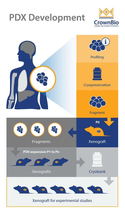
For the first implantation, patient-derived tumors may be implanted into immunodeficient mice either in the form of small tumor fragments or as tumor slurries derived from the blood of patients. The principal determinants of successful tumor engraftment into immunodeficient mice are the viability and sterility of the human tumor.

Cancer cells or tissues can be mixed with basement membrane matrix proteins (Matrigel®) before being injected into recipient animals, which enables the development of tumors with greater take rate and growth.

The time required to establish a PDX model varies by tumor type and grade. It can take anywhere from months to more than one year to finally get a model established. The first passage is the most difficult; if the tumor successfully grows, it will likely continue to grow in successive passages.

Orthotopically-Established PDX Better Mimic Patient Tumors

PDX can be implanted subcutaneously or orthotopically. PDX models are commonly established at subcutaneous sites, as cell implantation methods are easier and allow accurate monitoring of tumor size.

Orthotopically-implanted tumor cells have been shown to better mimic the drug response, growth patterns, and metastatic features of corresponding patient tumors, likely because of critical influence by the local stroma. Orthotopic implantation is more technically challenging and time-consuming, however, and often requires imaging (e.g. ultrasound, CT scan, etc.) or exploratory laparotomies to confirm the presence of internal tumors.

Validating PDX Models

One of the main laboratory management challenges with PDX models is drift from the original patient material. In order to ensure that the model is consistent during passages and over time, stringent validation and quality control is important. This is usually performed via:

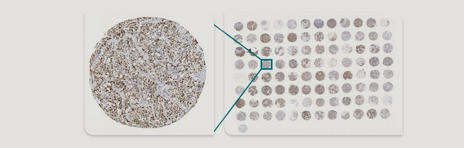
  • Pathology QC to confirm cancer type and to compare each PDX tumor to the original patient sample.
  • Genetic fingerprinting via STR genotyping.
  • Monitoring robust and consistent growth over multiple passages.

Humanized PDX Models in Immuno-Oncology

Humanized PDX models pair human tumors within a human immune system, more accurately reflecting human immunity and pathology. Researchers can use these models to assess the efficacy of immunotherapies (or combination immunotherapy regimens). There are different ways to create humanized PDX models, and humanized mice are available from a range of vendors which support engraftment and growth of PDX models.

The most common method for the generation of humanized mice is to transplant CD34-positive human hematopoietic stem cells (HSCs), isolated from the umbilical cord blood, into immunodeficient mice.

Humanized PDX models support multiple applications, including combination therapy studies with human-specific antibodies and immune targets, or evaluation of diversity across a population.


Related Posts