 Patient-derived xenografts (PDX) are the most predictive and translational xenograft models available for preclinical oncology research. However, by their nature, they are restricted to in vivo use and are often too costly and not amenable for platforms such as high throughput drug screening. This blog post looks at the in vitro and ex vivo assays that are being developed from PDX to overcome limitations and expand model use throughout the preclinical research spectrum.
PDX-Derived Cell Lines and Predictive In Vitro Research
The mainstay use of PDX models is in an in vivo, immunodeficient setting for agent efficacy assessment, most commonly executed as a “classical” xenograft study or in a Mouse Clinical Trial. With the improved predictability of PDX over conventional xenografts, and the unique, highly patient-relevant genetics (e.g. mutations, fusions) of some models, it would be useful to also use them in in vitro early stage drug development assays.
To this end, PDX-derived cell lines have been developed to provide a primary cell line for recapitulating clinically relevant features of human disease. Using these cell lines allows more predictive in vitro assays and a rapid transition to associated xenografts for PK/PD studies and parental PDX models for efficacy studies.
Parental PDX Features Maintained for a Range of In Vitro Assays
PDX-derived cell lines are developed from mouse stromal cell-depleted cancer cell cultures taken from PDX models. These undergo adaptation to grow within the in vitro/tissue culture setting as cell lines. PDX-derived cell lines are early passage (<10), to avoid drift from disease and maintain the essential histopathological features and genetic profiles of the original patient tumors/parental PDX models. Conserved features include:
- Mutational status (e.g. rare fusions or disease subtypes).
- Biochemical signaling.
- Response to anticancer targeted therapeutics.
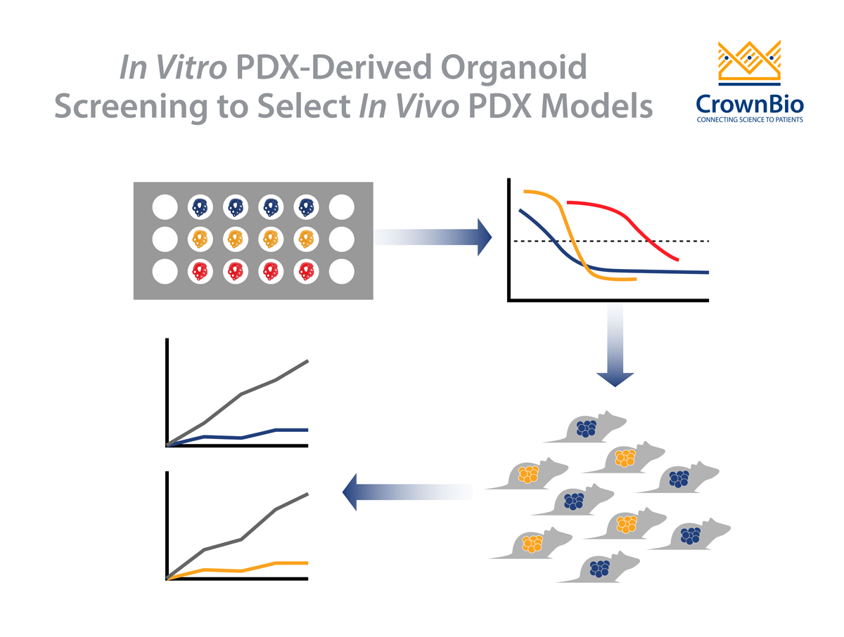
Fidelity between the parental PDX model and downstream cell lines has been confirmed via correlation of in vivo PDX tumor growth inhibition to in vitro cell line potency for targeted agents, dependent on the specific mutational background of the models.
Once developed, PDX-derived cell lines can be used as any standard in vitro cancer cell line resource – treated with novel agents and assayed in a variety of ways. The oncology and immuno-oncology research applications are wide-ranging, with assays providing data from simple potency evaluations to combination synergistic effects and cancer cell/immune cell interactions for I/O.
Ex Vivo PDX Support High Throughput Drug Testing
The use of the term “ex vivo” can often cause confusion compared with in vitro. In vitro is usually fairly well understood as referring to living things in culture, such as a cancer cell line grown in tissue culture. Ex vivo refers to things that were once living in vivo; e.g. cells which were growing in vivo and transferred to short-term culture for testing.
Taking PDX ex vivo supports powerful applications, such as high throughput assays, while maintaining the PDX advantage of predictability. These ex vivo models are usually used to assess the cytotoxicity or proliferation of cancer cells. Historically, this used simple clonogenic assays, but is now moving forward to higher throughput 3D screens, and more complex assays to more closely mimic the human setting e.g, the 3D Tumor Growth Assay (TGA).
Conserve the PDX Advantage for Compound Screening
PDX ex vivo 3D assay formats combine rapid compound testing and screening with PDX genomic analysis, thereby providing genetic mutation information to better interpret screening panel results.
These screens can be performed with frozen ex vivo tumor cells dissociated from freshly isolated early passage PDX tumor tissues. Using frozen cells helping to avoid lag times while waiting for PDX to grow.
In this setting, large panels of compounds are rapidly assessed across models which conserve all the advantages of PDX population heterogeneity and patient fidelity, alongside retention of human-derived stromal elements (which can be lost over PDX passage), lower costs, and faster study timelines. Conservation of PDX model characteristics and response are validated by comparison with matched in vivo data, which has been used to confirm the reliability of this screening platform.
Humanized and TIME-Aligned 3D TGA Better Represents Human Condition
Another new innovative ex vivo screening platform is the 3D TGA. The 3D assay is “humanized” and TME-aligned to investigate factors affecting drug response. This is achieved through the addition of hMSCs and cancer associated fibroblasts, which provide the paracrine signaling present in the solid tumor microenvironment. Hormones, glucose, and pH level are also controlled.
Similar to the ex vivo screen above, 3D TGA potency results have been correlated with parental PDX in vivo response to validate the platform. Relative TGIs between specific models treated with standard of care/targeted agents correspond to relative potencies ex vivo. Importantly, addition of cancer-associated fibroblasts to the 3D TGA has been shown to dampen ex vivo response (specifically for erlotinib in a lung cancer model), showing potential interaction between the fibroblasts (or their secreted factors) and cancer cell response. This highlights the importance of recapitulating the human condition in ex vivo assays.
In Vitro/Ex Vivo Assays Expand PDX Use in Preclinical Research
In vitro and ex vivo assay platforms benefit from the translational features of PDX, opening up powerful new options in early stage drug discovery while overcoming the inherent limitations of in vivo PDX tumor models.


