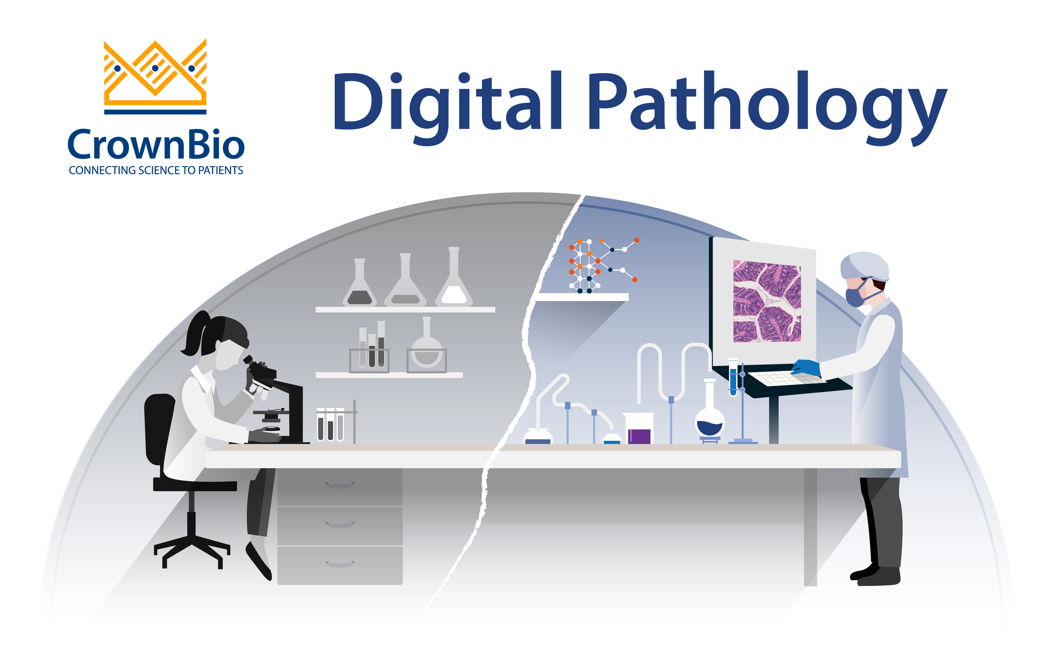
New technologies and methods are continuing to drive innovation in healthcare, including in the field of pathology. While a pathology report is generally considered the gold-standard for the diagnosis of many diseases, it’s commonly recognized that there is variability in the interpretation of tissue samples by pathologists. This is mostly due to human error and subjectivity, and consequently leads to inaccurate conclusions for patients.
To overcome this challenge, modern pathology practices are moving to an increasingly digital workflow. This incorporates tools for pathologists that are used to reduce workload, human error, and subjectivity. This then enables better conclusions to be made regarding diagnostic, prognostic, and predictive factors (such as biomarkers) associated with disease, especially cancer.
In this post, we explore what digital pathology is, and how it is being used for biomarker exploration and discovery, especially in the context of immuno-oncology (I/O).
Digital Pathology and Its Applications
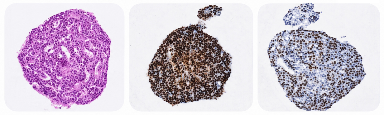
At the broadest level, the concept of digital pathology includes the capture, sharing, and interpretation of pathology information using digital technologies. The process begins by digitizing glass slides via a scanning device which generates a high-resolution image that is displayed on a computer screen.
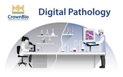
Subsequently, trained artificially intelligent (AI) algorithms, which rely on the use of machine learning (ML) and/or deep learning (DL), are used to assist a pathologist’s work by analyzing the digital slides at high‐resolution and high speed in an objective manner. AI algorithms are capable of quantifying several common metrics including:
Total biomarker stain area
Cellular-level biomarker positivity
Sub-cellular biomarker localization
Biomarker co-expression
Object quantification (e.g. microvessels)
Tissue classification
Cell-density, biomarker/cell proximity/distance analysis, and nearest-neighbor distances
Furthermore, image search capabilities represent a major benefit of digital pathology workflows. The ability to search images to match new patient pathology with previously diagnosed and curated cases offers improved diagnostic accuracy through the computational “consensus” of a primary diagnosis.
Below we describe how digital pathology is being used for the diagnosis and prognosis of diseases, and for biomarker research.
Diagnosis
One of the most widely used application for digitized pathology slides is to enhance a pathological diagnosis. Diagnostic AI algorithms search for shapes, features, or patterns utilizing image analysis. In some instances, AI has performed equally or better than human pathologists, including in cancer diagnoses that are complex and rely on grading schemes which are different than diagnoses based on tumor detection which is for the most part a binary decision (i.e. based on the presence or absence of cancer cells in tissue samples).
Overall, advances in AI have paved the way for new tools that can supplement a pathologist’s work, making their work more accurate and less burdensome.
Prognosis
Prognostic AI algorithms can play an important role in patient prognosis. For instance, specific biomarker assessment, such as Ki67 antigen in some cancer types, is important since it is correlated to the course of the disease. Investigators have devised amazingly accurate algorithms to quantify Ki67 stained tissue datasets, which are used to reduce the pathologist’s workload while performing equally or better.
Prognostic algorithms are also being leveraged to evaluate images from large datasets to discover novel predictive and prognostic parameters that correlate with the clinical course of disease.
Biomarker Analysis
Digital pathology is a valuable tool for biomarker exploration and discovery. For instance, AI algorithms are enabling researchers to analyze the density and distribution of immunohistochemically and/or fluorescently labeled biomarkers within large datasets of tissue samples.
These data can then be correlated to different parameters with the goal of stratifying patients to:
Develop precision medicine approaches including companion diagnostics
Develop better prognostic models
Provide insights into drug resistance, among other possibilities
Overall, digital pathology is providing novel opportunities for the discovery and utilization of biomarkers that were not previously possible.
Digital Imaging Platforms for Biomarker Assessment
A variety of digital imaging platforms are available for quantitative biomarker evaluation, each with its own set of capabilities. Some of these solutions are listed below:
Ventana Companion Algorithm to evaluate breast panel biomarkers (e.g. ER, PgR, HER2, Ki67, P53) and support antibody assays.
Tissue Phenomics® by AstraZeneca was introduced to clinical programs in I/O to support biomarker identification.
Aperio Digital Pathology by Leica Biosystems is a platform that integrates a digital microscope with image analysis software.
HALO by Indica laboratories offers immunohistochemistry and fluorescence modules of quantitative tissue analysis which are designed primarily for research purposes.
QuantCenter by Sysmex is designed for digital whole slide quantification processes and provides several modules for tissue classification, immunohistochemical quantification, and molecular pathology.
Cognition Master Professional Suite by VMscope can be integrated into laboratory information management system and offers modules for scoring of several biomarkers (e.g. Ki67, ER, PgR, CD3/4/8/15/20), tumor infiltrating lymphocytes, and vascular stenosis.
TissueGnostics provides image analysis solutions for clinical and research evaluation of biomarkers in breast cancer.
VirtualDualStaining™ by Visiopharm aligns a pancytokeratin-stained consecutive section of the tumor with the immunohistochemically stained biomarker under investigation, allowing automated identification of tumor regions.
In addition to these commercial platforms, there also exist open‐source platforms. One of the first for image analysis was a tool developed by the NIH known as ImageJ. This provides morphological parameters and is widely used for biomedical image analyses.
Another open-sourced platform known as CellProfiler provides supervised machine learning‐based classification to perform imaging‐based diagnoses. Finally, QuPath has a special focus on digital pathology and whole slide image analysis and offers unsupervised machine learning‐based cell detection and supervised classification of whole slide images, tumor identification, and high‐throughput biomarker evaluation. This tool has been embraced by the research community since it was first published in 2017, and has been heavily cited in peer‐reviewed publications.
Digital Pathology in Immuno-Oncology
The coincidental rise of both I/O and AI has led to a renewed and strong interest in digital pathology, which is highly applicable to I/O biomarker exploration and discovery studies. The pathological signatures defining the tumor-immune microenvironment are highly complex, with protein, cellular, and/or morphometric markers. This is beyond what a typical human pathologist can reasonably analyze, especially when faced with a daunting volume of slides.
By using a digital pathology approach, analyses are performed efficiently across entire slides and/or within regions of interest, in absolute or relative terms, and within a single slide or across multiple sequential sections. A digital approach also allows for multi-omic analyses, with some platforms providing a common format of target-specific bar-coded labels for both proteins and nucleic acids, which allow for computational analyses for deconvolution and quantification of targets.
Interestingly, I/O is also driving new clinical applications for digital pathology. For example, assessment of PD-L1 to guide treatment with checkpoint inhibitors (i.e. selecting patients more likely to respond to therapy) is becoming increasingly complex given the many different factors that can influence its expression. These include the type of assay and antibody clone used, the scoring cut-off, previous drugs taken by the patient, and the inherent nature of the sample itself, among other possible factors.
Against this background, digital pathology offers the ability to automate these analyses with the goal of reducing subjectivity and human error. Future advances in digital pathology may include the compilation of clinically relevant information from stained slides to develop companion diagnostics without needing to validate new biomarkers or assays.
Bolstered by the same advances in I/O and AI, and developments in digital pathology, multiplex spatial tissue analysis (MSTA) allows for the detailed evaluation of the tumor microenvironment (TME). MSTA refers to methods used for assessing the specific location of multiple biomarkers (protein and/or nucleic acid markers) within a tissue sample. This has typically been limited to 3 – 5 protein markers for immunohistochemistry and 7 – 9 markers for immunofluorescence.
Novel methodologies - such multi-cycle stain stripping/quenching/ recycling methods, photo cleavable barcodes and spatially defined UV exposure, or laser/ion-beam ablation of metal-or isotope-labelled antibodies) - coupled with digital approaches have unleashed the potential of MSTA. The technique is now able to simultaneously evaluate dozens to more than 1,000 nucleic acid and/or protein markers (in regions of interest) while maintaining some of the spatial context. Some platforms can even assess spatial whole RNA transcriptomes at the near-single cell level.
There are numerous providers of MSTA platforms, which differ from product-to-product in terms of specifications and capabilities. Some of the platforms and their manufacturers include (not a complete list):
GeoMx DSP (NanoString)
MultiOmyx (NeoGenomics)
Phenoptics, CODEX (Akoya Biosciences)
MACSima (Miltenyi)
Cell DIVE (GE Life Sciences)
RNA-Scope (Advanced Cell Diagnostics)
UltraPlex (Cell IDx)
Zellkraftwerk (ChipCytometry)
Currently, the use of MSTA is primarily focused on translational research (e.g. biomarker discovery, pharma R&D, clinical trials) and potential clinical applications (e.g. clinical and companion diagnostics). A recent systematic review and meta-analysis published in JAMAOncology provides compelling evidence that the use of multiplex immunohistochemistry/ immunofluorescence had a better diagnostic accuracy for the assessment of response to PD-1/L1 checkpoint blockade compared to other companion diagnostic types such as tumor mutational burden (TMB), gene expression profiling (GEP), single-plex PD-L1 IHC, and composites of these methods.
Conclusion
Digital pathology is playing an increasingly important role in the diagnosis, prognosis, and identification of predictive factors, such as biomarkers, associated with disease, especially cancer. While digital pathology represents a novel tool to benefit the work of pathologists, it is also enabling better exploration and discovery of biomarkers which are key for more widespread adoption of personalized medical care which can improve patient outcomes.
Cite this Article
Bourré, L., (2020) Digital Pathology - Crown Bioscience. https://blog.crownbio.com/digital-pathology
New data from translational research groups, platform developers, and earlystage biotech teams point to a clear trend in 2026: scalable 3D assays and …
Immunooncology (IO) drug development increasingly depends on understanding human specific immune mechanisms. However, these subtle, highly coordinated…
An Interview with Crown Biosciences' SVP of Commercial, Alex Slater Meet Alex Slater, Senior Vice President of Commercial at Crown Bioscience. With a …