<img height="1" width="1" src="https://www.facebook.com/tr?id=1582471781774081&amp;ev=PageView &amp;noscript=1">
  • Menu
  • crown-logo-symbol-1-400x551

Find it Quickly

Get Started

Select the option that best describes what you are looking for

  • Services
  • Models
  • Scientific Information

Search Here For Services

Click Here to Start Over

Search Here For Models

Click Here to Start Over

Search Here For Scientific Information

Click Here to Start Over

In Vitro

Boost oncology drug discovery with XenoBase®, featuring the largest cell line selection and exclusive 3D organoid models. Benefit from OrganoidXplore™ and OmniScreen™ for rapid, in-depth analysis.

Learn More

In Vivo

Enhance drug development with our validated in vivo models, in vitro/ex vivo assays, and in silico modeling. Tailored solutions to optimize your candidates.

Learn More

Tissue

Experience ISO-certified biobanking quality. Access top biospecimens from a global clinical network, annotated by experts for precise research.

Learn More

Biomarkers and Bioanalysis

Leverage our global labs and 150+ scientists for fast, tailored project execution. Benefit from our expertise, cutting-edge tech, and validated workflows for reliable data outcomes.

Learn More

Data Science and Bioinformatics

Harness your data and discover biomarkers with our top bioinformatics expertise. Maximize data value and gain critical insights to accelerate drug discovery and elevate projects.

Learn More

KRAS

Accelerate innovative cancer treatments with our advanced models and precise drug screening for KRAS mutations, efficiently turning insights into clinical breakthroughs.

Learn More

EGFR

Advance translational pharmacology with our diverse pre-clinical models, robust assays, and data science-driven biomarker analysis, multi-omics, and spatial biology.

Learn More

Drug Resistance

Our suite integrates preclinical solutions, bioanalytical read-outs, and multi-omics to uncover drug resistance markers and expedite discovery with our unique four-step strategy.

Learn More

Patient Tissue

Enhance treatments with our human tumor and mouse models, including xenografts and organoids, for accurate cancer biology representation.

Learn More

Bioinformatics

Apply the most appropriate in silico framework to your pharmacology data or historical datasets to elevate your study design and analysis, and to improve your chances of clinical success.

Learn More

Biomarker Analysis

Integrate advanced statistics into your drug development projects to gain significant biological insight into your therapeutic candidate, with our expert team of bioinformaticians.

Learn More

CRISPR/Cas9

Accelerate your discoveries with our reliable CRISPR solutions. Our global CRISPR licenses cover an integrated drug discovery platform for in vitro and in vivo efficacy studies.

Learn More

Genomics

Rely on our experienced genomics services to deliver high quality, interpretable results using highly sensitive PCR-based, real-time PCR, and NGS technologies and advanced data analytics.

Learn More

In Vitro High Content Imaging

Gain more insights into tumor growth and disease progression by leveraging our 2D and 3D fluorescence optical imaging.

Learn More

Mass Spectrometry-based Proteomics

Next-generation ion mobility mass spectrometry (MS)-based proteomics services available globally to help meet your study needs.

Learn More

Ex Vivo Patient Tissue

Gain better insight into the phenotypic response of your therapeutic candidate in organoids and ex vivo patient tissue.

Learn More

Spatial Multi-Omics Analysis

Certified CRO services with NanoString GeoMx Digital Spatial Profiling.

Learn More

Biomarker Discovery

De-risk your drug development with early identification of candidate biomarkers and utilize our biomarker discovery services to optimize clinical trial design.

Learn More

DMPK Services

Rapidly evaluate your molecule’s pharmaceutical and safety properties with our in vivo drug metabolism and pharmacokinetic (DMPK) services to select the most robust drug formulations.

Learn More

Efficacy Testing

Explore how the novel HuGEMM™ and HuCELL™ platforms can assess the efficacy of your molecule and accelerate your immuno-oncology drug discovery programs.

Learn More

Laboratory Services

Employ cutting-edge multi-omics methods to obtain accurate and comprehensive data for optimal data-based decisions.

Learn More

Pharmacology & Bioanalytical Services

Leverage our suite of structural biology services including, recombinant protein expression and protein crystallography, and target validation services including RNAi.

Learn More

Screens

Find the most appropriate screen to accelerate your drug development: discover in vivo screens with MuScreen™ and in vitro cell line screening with OmniScreen™.

Learn More

Toxicology

Carry out safety pharmacology studies as standalone assessments or embedded within our overall toxicological profiling to assess cardiovascular, metabolic and renal/urinary systems.

Learn More

Preclinical Consulting Services

Learn more about how our consulting services can help to support your journey to the clinic.

Learn More

Our Company

Global CRO in California, USA offering preclinical and translational oncology platforms with high-quality in vivo, in vitro, and ex vivo models.

Learn More

Our Purpose

Learn more about the impact we make through our scientific talent, high-quality standards, and innovation.

Learn More

Our Responsibility

We build a sustainable future by supporting employee growth, fostering leadership, and exceeding customer needs. Our values focus on innovation, social responsibility, and community well-being.

Learn More

Meet Our Leadership Team

We build a sustainable future by fostering leadership, employee growth, and exceeding customer needs with innovation and social responsibility.

Learn More

Scientific Advisory Board

Our Scientific Advisory Board of experts shapes our strategy and ensures top scientific standards in research and development.

Learn More

News & Events

Stay updated with Crown Bioscience's latest news, achievements, and announcements. Check our schedule for upcoming events and plan your visit.

Learn More

Career Opportunities

Join us for a fast-paced career addressing life science needs with innovative technologies. Thrive in a respectful, growth-focused environment.

Learn More

Scientific Publications

Access our latest scientific research and peer-reviewed articles. Discover cutting-edge findings and insights driving innovation and excellence in bioscience.

Learn More

Resources

Discover valuable insights and curated materials to support your R&D efforts. Explore the latest trends, innovations, and expertly curated content in bioscience.

Learn More

Blogs

Explore our blogs for the latest insights, research breakthroughs, and industry trends. Stay educated with expert perspectives and in-depth articles driving innovation in bioscience.

Learn More

  • Platforms
  • Target Solutions
  • Technologies
  • Service Types

Publication Spotlight: DSP vs. Visium: A Comparative Study in Spatial Transcriptomics for Cancer Research

Dr. Alexander Swarbrick's recent paper on bioRxiv, titled "An experimental comparison of the Digital Spatial Profiling and Visium spatial transcriptomics technologies for cancer research" delves into a comparative analysis of the DSP and Visium platforms. While both technologies displayed commendable prowess in spatially profiling breast cancer tissues, they shine in distinct areas of application.

The Complex Landscape of Tumors: A Call for Advanced Spatial Omics Tools

 Tumors are far from simple accumulations of cancer cells. Instead, they are intricate ecosystems teeming with various cellular subtypes. The spatial organization of these cells within tumors offers a window into the molecular intricacies of cancer, influencing tumor progression and response to treatments. Traditional methods, like multiplexed immunofluorescence, can only probe so deep. The emerging realm of spatial omics technologies promises to bridge this gap. Among these, the NanoString GeoMx® Digital Spatial Profiling (DSP) and 10x Genomics Visium platforms are turning heads, empowering researchers to gain a comprehensive understanding of tumor heterogeneity.

Crown Bioscience's Certified
NanoString Spatial Multiomics
Services

Learn More


The Comparative Dive: DSP vs Visium Platforms

Experimental Setup

For a rigorous comparison between DSP and Visium, the researchers devised an intricate experimental setup, using four primary breast cancer tissue samples and two cultured cell lines (Figure 1). This approach ensured a detailed exploration of the diverse cellular interactions characteristic of tumors.

Figure 1: Experiment overview.
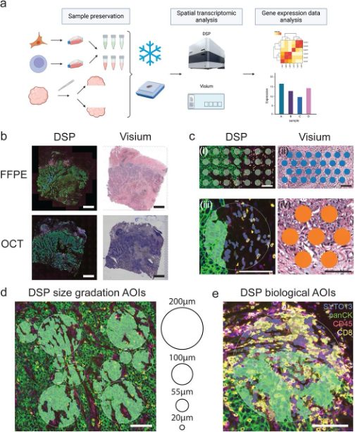
(a) Schematic illustration of sample preservation and experiment workflow. Cultured Jurkat and SKBR3 cells were mixed at six different ratios and preserved as OCT or FFPE samples. Tissue samples were sliced in the middle and the resulting two pieces were preserved as OCT or FFPE samples respectively. Closest possible sections were used for DSP and Visium assays. The illustration was created with BioRender.com. (b) Example images of tissue morphology. FFPE and OCT sections from sample 4806 were processed for DSP or Visium assays. Note the overall matching morphology between FFPE and OCT samples and between sections used for DSP and Visium assays. Scale bars = 1mm. (c) Example of DSP AOIs and Visium spots used for direct comparison. (i) Example of Visium mimic AOIs across the tumour-stroma interface. (ii) Example of Visium spots across the tumour-stroma interface used for direct DSP and Visium comparison. (iii) Example of segmented comparison DSP AOIs. Each AOI was segmented into pan-cytokeratin positive and pan-cytokeratin negative segments according to the immunofluorescence signal. (iv) Example of Visium spots located in the matching region where segmented comparison DSP AOIs were collected. Scale bars = 100μm. (d) Example of size gradation and biological DSP AOIs to test the performance of segmentation of the DSP platform. Scale bars = 100μm.
 

Key Observations

  • Gene Expression Correlation: High correlation was observed in controlled cell array samples between the two platforms. However, the complex nature of breast cancer tissues revealed nuanced differences in their gene detection capabilities.
  • Diving Deep into Tissue Analysis: While both platforms displayed an impressive depth in tissue analysis, Visium on OCT processed samples lagged slightly in gene detection compared to its FFPE counterpart. The nuances in tissue preparation significantly impact the outcomes.

Spotlight on Platform Strengths

  • Visium by 10x Genomics: With its unbiased approach, Visium offers a comprehensive landscape of entire tissues. This bird's-eye view allows researchers to unearth spatial heterogeneity in unmatched detail. Visium excels in initial exploratory research phases, offering whole transcriptomes across expansive tissue areas.
  • NanoString GeoMx DSP: DSP, on the other hand, excels in precision. Its unique ability to deep-dive into specific regions of interest, focusing on intricate molecular profiling, makes it an invaluable tool for hypothesis-driven research.


Exploring Specific Regions with DSP

The DSP platform's prowess was further showcased when zooming into granular regions. This deep dive revealed the intricate interplay between closely associated cell types like CD8 T cells and surrounding cells (Figure 2). However, ensuring purity in transcriptomic profiles amidst such tight cellular interactions remains a challenge.

Figure 2: Segmentation by the DSP platform allow profiling of more specific gene expression features.
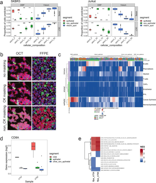
(a) Predicted proportion of SKBR3 and Jurkat cells in segmented DSP AOIs and Visium spots in cell array samples. (b) Example of segmented DSP AOIs on cell array samples. All cells were labelled with SYTO13 for nuclei stain. Jurkat and SKBR3 cells were labelled with anti-CD45 (red) or pan-cytokeratin (green) antibodies respectively. Epithelial or non-epithelial DSP AOIs were then sampled based on the fluorescent signals in a sequential manner. (c) Predicted proportion of cancer and TME cell types in segmented DSP AOIs and Visium spots in tissue samples. (d) Normalised expression of CD8A in CD8 and non CD8 segments in DSP data. (e) NES of top 5 significantly dysregulated GOBP pathways identified between CD8 segments and adjacent non-CD8 TME segments by the DSP platform. Significance threshold was set as q < 0.25.


Conclusion

The world of spatial transcriptomics isn't one-size-fits-all. While both DSP and Visium are powerhouses in their own right, the choice boils down to the specific research question at hand. This comparative analysis paves the way for informed decisions, enabling researchers to harness the full potential of spatial transcriptomics.

Crown Bioscience's Certified
NanoString Spatial Multiomics
Services

Learn More


Related Posts