Leveraging Targeted Mouse I/O RNA-Seq Panels to Improve the Translatability of Immuno-Oncology Studies
11:54
SHARE
In this post, we explore how a targeted RNA-Sequencing (RNA-Seq) panel can be incorporated into preclinical drug development to evaluate the impact of immune effectors in mediating anti-tumor immunity of novel immuno-oncology (I/O) agents. A case study highlights how RNA-Seq panels can complement traditional protein-based assays to gain a truly comprehensive picture of novel immunotherapies.
Precision Medicine: The Promise and the Challenges
Checkpoint blockade and other forms of cancer immunotherapies can induce durable clinical responses for a growing number of cancer indications. Remarkably, such responses are being observed in advanced cancer types for which effective therapies were previously not available.
However, similar to other anti-cancer treatments, only a subset of patients benefits from therapies that enhance anti-tumor immunity. This has led to an urgent need to identify novel biomarkers that can identify patients most likely to respond to immunotherapy.
Such biomarkers should ideally involve features of the tumor microenvironment (TME) since the composition and density of immune cells in the TME is well known to profoundly influence tumor progression, and efficacy of anti-cancer therapies.
Assays that allow for the genomic profiling of the TME are playing an increasingly important role in improving current and novel immunotherapy regimens.
Assays for TME characterization
High-throughput RNA-Seq has revolutionized cancer genomics and represents a promising avenue for accessing the complex molecular features and interactions of the TME.
Until now, protein-based methods such as fluorescence-activated cell sorting (FACS), immunohistochemistry (IHC) and immunofluorescence (IF) have been considered the gold standards in estimating the immune cell content within a sample.
While these techniques can still provide valuable insights, these methods have technical limitations that prevent them from being more broadly applicable. For instance, a continuous challenge is the availability, or lack thereof, of high-quality validated antibodies targeting potential protein biomarkers.
Further, IHC and IF, and to a lesser degree FACS, are limited in the number of markers that can be examined simultaneously (See Table 1 for some common limitations associated with the different methods).
Table 1. Methods used for the quantitative analysis of the TME.
Method
Limitations
FACS
Large amount of material needed and limited number of markers
IHC / IF
Not representative if the tumor is heterogeneous and limited number of markers
RNA-Seq
Provides no information on cellular composition
single-cell RNA sequencing (sc-RNAseq)
Expensive and bias due to tissue dissociation
Next-generation RNA-Seq is a powerful high-throughput substitute or complementary method to protein-based technologies and provides an accurate measure of gene expression across the entire genome without requiring the pre-selection of specific targets.
Moreover, the capacity to obtain both expression quantification and nucleic acid sequence information from RNA-Seq can be harnessed to measure immunological signals that are often obtained through multiple, distinct technologies.
With progress in sc-RNAseq, the complex interplay of tumor cells with the TME can now be reconstructed with great precision. These benefits can reduce costs and allow researchers to meet or fast-track project deadlines.
The Need for a Murine Next-Generation Sequencing (NGS)-Based Assay as a Basic Tool for Preclinical Drug Development
The prognostic value of RNA-based molecular features describing the TME has been demonstrated in recent clinical trials, where gene expression signatures for tumor infiltrating immune cells have been generated for several malignancies.
But this type of evaluation does not have to be restricted to the clinical phase of drug development.
Instead, a better understanding of the role the TME plays in an immunotherapy’s mechanism-of-action (MoA) and efficacy should ideally be developed before moving to human clinical studies.
By using assays that provide insights into the TME earlier in the drug development process, and across a range of preclinical models suitable for I/O studies, we aim to improve patient stratification to enhance both efficacy and safety of a novel agent.
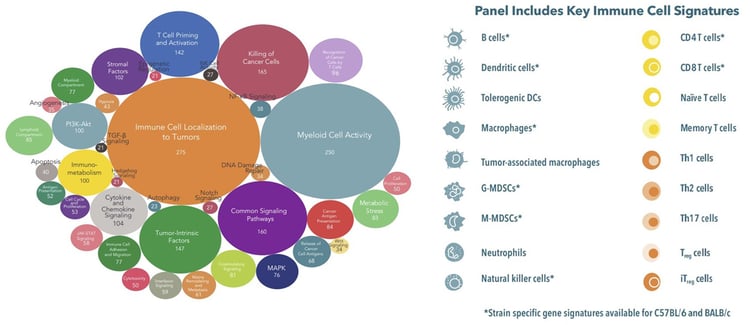
Mouse Immuno-oncology targeted RNA-Seq panels (known as Mouse I/O RNA-Seq Panels) are available to evaluate genes that are associated with tumor immunity. This enables rapid transcriptomic insights into key immune cell populations and I/O pathways and processes in the TME (Figure 1).
For instance, in a Mouse I/O RNA-Seq available panel that encompasses 1,080 genes, immune cell signatures, surface markers, transcriptomic immune cell-specific biomarkers, and key pathways at the interface of the tumor, TME, and immune response can all be interrogated.
Figure 1. Characteristics of the Mouse I/O RNA-Seq panel
A typical workflow for a mouse I/O RNA-Seq panel should feature a simple design and offer flexibility in the source and type of study samples that can be analyzed (Figure 2).
Having flexibility in the workflow is important so that samples (e.g., extracted RNA, frozen tissue, whole blood, immune cell pellets) generated through syngeneic and Tumor homograft model studies, or from in-house studies, or externally generated samples, can seamlessly be integrated into the workflow.
Finally, the final output should include both raw and normalized data as well as a comprehensive report with multiple group/condition comparisons.
By using a collection of validated syngeneic and tumor homograft models that cover a range of cancer types and immune profiles, large-scale screening studies allow researchers a unique opportunity to:
Gain deep insights into an agent’s MoA
Identify biomarkers that may predict response
Use relevant preclinical models with human relevant cancers
Figure 2. Features of the Mouse I/O RNA-Seq panel workflow.
RNA-Seq platforms have several advantages over other commonly used transcriptomic technologies (Table 2 shows a comparison of these two assays).
A comparison study of RNA-Seq with NanoString (a variation of DNA-based microarray technology) showed the mouse I/O RNA-Seq panel had higher accuracy and sensitivity, and the ability to discriminate between different mouse strains and carried shorter turnaround times rendering it more cost effective.
Table 2. Platform Comparison: mouse I/O RNA-Seq panel vs microarray based I/O profiling
Assay Comparison
Mouse I/O RNA-Seq Panel
Microarray I/O Profiling
Technology
Targeted deep NGS
Target mRNA (cDNA) hybridize with DNA probes
Target Molecules
mRNAs
mRNAs
Throughput
High (1080 genes)
High (hundreds to thousands of genes, depending on vendor)
Accuracy
High
Low-medium
Sensitivity on detecting low expressing genes
High
Low-medium
Mouse strain discrimination
Yes
No
Turnaround time
2-3 weeks
3-10 weeks depending on vendor
Cost
Low
Medium
Mouse I/O RNA-Seq panels can complement and enhance fluorescence-activated cell sorting (FACS)-based I/O profiling
As described above, RNA-Seq overcomes some key limitations of protein-based methods for TME profiling. Along with differences in target molecules, employing a mouse I/O RNA-Seq panel has much higher throughput compared to FACS (up to 1080 targets versus ~30), increased accuracy, and is suitable for large scale sample screening.
In addition, mouse I/O RNA-Seq panels can be used as a discovery tool while FACS is best suited for hypothesis testing and validation.
While a recognized limitation for RNA-Seq panels is the slower turnaround time as compared to some protein-based methods, RNA-Seq platforms do deliver more comprehensive sample analyses with comparatively lower costs on a per sample basis (see Table 3 for RNA-Seq Panel benefits).
Further, it is worth noting that Mouse I/O RNA-Seq platforms do not necessarily replace protein-based profiling methods, but they should be viewed as complementary for obtaining the most complete picture of immune cell content and effector pathway activation within tumor samples. This power of this complementary approach is highlighted in the following case study.
Table 3. Summary of Mouse I/O RNA-Seq Panel Benefits
Mouse I/O RNA-Seq Panel Benefits
Comprehensive
Expression level of 1080 genes from a single sample
Wide coverage in tumors, TME, and immune response
Flexible Sample Types
Extracted RNA
Multiple tissues: tumor, blood, immune cells etc.
High-Throughput
Suitable for large-scale immunotherapy screening, as well as examining multiple tissues in parallel
High Sensitivity High Accuracy
>3 times the sequencing depth of RNA-Seq
Avoids biases on non-protein coding RNAs like mitochondria RNAs
Easy Integration
As standalone service or streamline with preclinical in vitro studies
Complementary with FACS, IHC and WB
Cost-Effective Fast Turnaround
Lower cost per sample
3-week turnaround time
Case Study: Determining the Response to Immunotherapy Under Varied Drinking Water pHs
Many studies have demonstrated that the gut microbiome (GM) plays an important role in immunotherapy response in both animal models and humans. In vivo preclinical variables, such as diet, housing, bedding and the pH of drinking water (such as acidified drinking water commonly used in research facilities), are known to influence the GM.
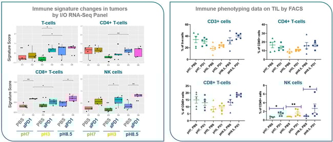
However, the effects of these variables on immunotherapy response remains unclear. This study therefore investigated the effects of drinking water pH on anti-PD-1 efficacy in MC38 murine colon cancer models using targeted RNA-Seq and FACS as complementary data sources.
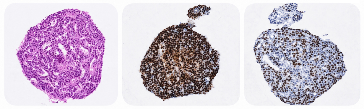
RNA-Seq analysis of MC38 tumors was consistent with FACS data, confirming that mice receiving acidified water had the lowest total lymphocyte infiltration (CD45+), T cells (CD3+, CD4+ and CD8+) and highest percentage of NK cells.
These results indicate that acidified water can modulate the response to immunotherapy by inducing an immunosuppressive TME (Figure 3).
Figure 3. Immune signature (RNA-Seq) and immune cell phenotyping (FACS) analysis of MC38 tumors following anti-PD-1 therapy and administration of drinking water with different pH levels.
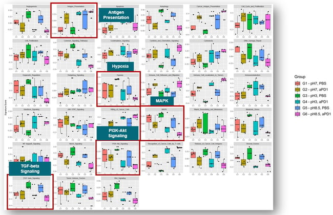
Leveraging the expanded number of immune signatures covered by a mouse I/O RNA-Seq Panel, pathway analysis revealed that hypoxia, MAPK and TGF-β signaling were also impacted by the pH, and alkaline conditions promoted antigen presentation.
Figure 4. Immune signature analysis of MC38 tumors treated with anti-PD-1 therapy followed by administration of drinking water a different pH levels.
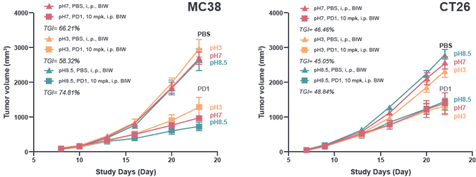
In the efficacy portion of the study, MC38 and CT26.WT colon cancer models were assessed. Different pH levels of drinking water were found to have a more pronounced impact on anti-PD-1 efficacy for the MC38 model.
Specifically, anti-PD-1 treatment in this model showed the largest tumor growth inhibition (TGI) with alkaline water (TGI=74%), followed by pH neutral water (TGI=66%) and then acidified water (TGI=58%).
Figure 5. Responses to anti-PD-1 treatment with drinking water at different pH levels of MC38 and CT26.WT murine colon cancer models.
In summary, pH neutral to alkaline water led to expansion of immune effector cells, restraining of tumor suppressive subsets and activation of signaling pathways that promote anti-tumor immunity.
Based on these data:
Alkaline water is recommended for preclinical I/O efficacy studies that employ syngeneic mouse models
Alkaline drinking water should also be provided when exploring therapies intended to enhance the gut microbiome
Acidified water should be avoided when conducting I/O studies as it potentially weakens the response to therapies by modulating the microbiome community
Conclusion
Leveraging RNA-Seq panels offers researchers a variety of unique benefits that complement traditional protein-based assays. Together, these assays can help pinpoint an MoA and identify predictive biomarkers that can possibly improve the translatability of a new immunotherapy agent.
By incorporating strategies early in development that can fully characterize the TME, researchers have the best opportunity to accelerate their immunotherapy studies and position them for clinical success.
For more information on our Mouse I/O RNA-Seq panel, view our latest on-demand webinar.
Cite this Article
Vallerand, D., (2021) Leveraging Targeted Mouse I/O RNA-Seq Panels to Improve the Translatability of Immuno-Oncology Studies - Crown Bioscience. https://blog.crownbio.com/leveraging-targeted-mouse-i-o-rna-seq-panels
New data from translational research groups, platform developers, and earlystage biotech teams point to a clear trend in 2026: scalable 3D assays and …
Immunooncology (IO) drug development increasingly depends on understanding human specific immune mechanisms. However, these subtle, highly coordinated…
An Interview with Crown Biosciences' SVP of Commercial, Alex Slater Meet Alex Slater, Senior Vice President of Commercial at Crown Bioscience. With a …