Cancer research studies have traditionally relied on 2D systems, but researchers are increasingly turning to 3D cell cultures. This is because they better recapitulate the original tissue architecture, which is known to affect key cellular processes, including cell signaling, proliferation, viability, and drug response. By providing more physiologically relevant models, 3D cell cultures can bridge the gap between in vitro and in vivo models.
Despite several options for 3D in vitro systems, the choice is not always straightforward. It largely depends on the biological question(s), such as understanding disease pathways, investigating mechanisms of action, or predicting patient response. Although spheroids have historically been the most common 3D models, more patient-relevant options have emerged, such as organoids.
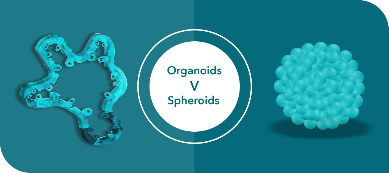
This blog post examines key differences between spheroid and organoid models, so that you can make the best choice for your specific needs.
Key Differences Between Organoids and Spheroids
Mechanism Driving the Formation of the 3D Structure. Spheroids are formed primarily via cell-to-cell adhesion (i.e., cell aggregates). The physical/chemical properties of the growing culture force the cells together to generate a 3D structure. Spheroids can be derived from immortalized cell lines, primary cells, or fragments of human tissue. In contrast, organoid formation is driven by the self-organizing properties of stem cells, which self-renew and differentiate /in vitro/, resulting in 3D structures containing multiple differentiated and physiologically functional cell lineages. As a result, organoids are more physiologically relevant and a more suitable option for cancer drug discovery applications because they produce data with greater translational potential to /in vivo/. They have been [shown to have high clinical relevance](https://www.science.org/doi/10.1126/science.aao2774) . Tumor organoids can be developed directly from primary tumor tissue (i.e., patient-derived organoids (PDOs)) or patient-derived xenograft (PDX) organoids (PDXOs).
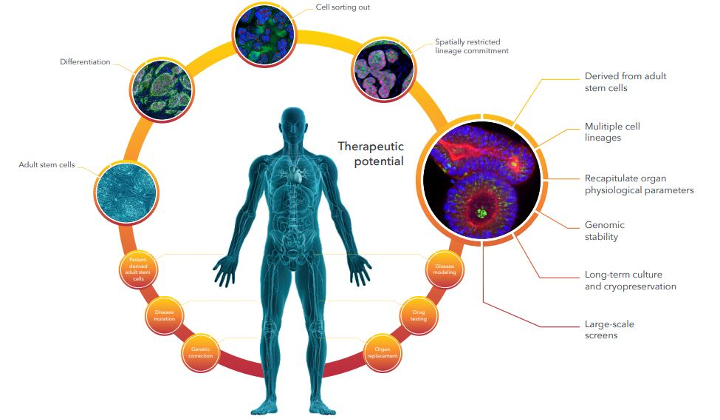
Figure 1. Generating Patient-Derived Organoids: A Comprehensive Approach.
Cellular Composition of the 3D Structures. Organoids have a multicellular identity that faithfully recapitulates the complexity of the original tumor (or organ). They develop this identity because of the stem cells that generate them and differentiate into multiple downstream and specialized progeny. Spheroids are often a monoculture and therefore commonly lack the multicellular identity of an in vivo tumor. This is because, similar to 2D cell lines, tumor spheroids are cultured in conditions that select for a high proportion of poorly differentiated cells with high replicative potential, which are generally more resilient and more capable of growing in vitro. These characteristics can affect drug response and produce data that poorly translate to the clinic. From a cancer drug discovery perspective, organoids’ multicellular identity means that they are more patient-relevant and predictive of patient response to drugs compared to tumor spheroids.
Culture Robustness. Tumor organoids developed using Hubrecht organoid technology (HUB) protocols have a preserved cancer stem cell (CSC) component and so can be propagated over long periods and even biobanked and resuscitated from frozen for follow-up studies while predictably retaining the phenotypic and genetic features of the parental tumor over multiple generations, including structural and cellular heterogeneity and somatic mutations.
Although tumor spheroids are used in in vitro drug screens, they are obtained with variable efficiency and have variable CSC levels. The literature also reports changes in spheroids’ response profile over time, making them a less robust system compared to organoids. Tumor spheroids derived from primary cells can recapitulate the genomic and multicellular profiles of their parental tumor tissue. However, they are considered a one-shot solution because of their very limited lifespan in culture; they contain a high proportion of differentiated cells and therefore can quickly become senescent. In addition, deriving primary cells from some cancer types remains notoriously challenging, and typical culture conditions often select for cells that can best withstand the in vitro environment.
Data Quality. If a multicellular mixture of cells, such as primary cells, is used to develop tumor spheroids, then readouts from classical assays will tend to have a greater signal-to-noise ratio compared to organoids, which consist of a pure epithelial compartment. A notable benefit of tumor spheroids, especially those derived from immortalized cell lines, is the wealth of historical data that can be used for comparison.
Cancer Drug Discovery Studies Using Organoids Versus Spheroids
Tumor-derived organoids have been extensively investigated for cancer drug discovery, including identifying targets, evaluating the efficacy of drug candidates, and interrogating novel combination strategies. The in vitro models are especially attractive because they can be more cost efficient compared to in vivo PDXs and allow for earlier decision-making instead of waiting for late-stage in vivo model data. Furthermore, they are adaptable to standard in vitro assays, such as IC50 measurements and luminescent cell viability (e.g., CTG) readouts for cell viability. They can also be combined with innovative high content imaging (HCI) and analysis (HCA) platforms to generate extremely detailed cellular physiology profiles of complex cellular systems, including multicellular structures. By doing so, researchers can significantly expand the amount of information obtained from cellular assays compared to fixed endpoint assays, such as CTG or nuclei count.
However, organoid applications in drug development have some key differences compared to spheroids. As noted, spheroids derived from primary cells directly from patients are not readily scalable, and samples can become limited as studies progress. Ideally, fresh tissue should be used for optimal translatability. If it becomes scarce, frozen samples can be used, if they were biobanked.
Although sourcing patient tumor tissue can be a challenge for developing PDOs, these organoids can be used to generate PDX models as a source of PDXOs that can then serve as an alternative and prolific option to establishing tumor organoids. This is because PDXOs have been shown to preserve the clinical relevance of PDOs yet provide additional operational simplicity for large-scale screens. For instance, Crown Bioscience has developed our Organoid Models Database, an extensive searchable biobank consisting of almost 350 tumor organoid models derived from its collection of PDX models with information on histopathology, IC50, genomic, and transcriptomic analysis data associated with drug response (plus an additional 200+ models from primary patient samples). By leveraging existing collections of well-characterized PDX models, it is feasible to generate large PDXO libraries that optimally capture the heterogeneity and diversity observed in patient populations.
Pairing in vitro models with matched in vivo models is not always possible with tumor-derived spheroids, which represents yet another advantage of organoids in drug discovery. Additionally, the organoid model offers the benefit of sourcing both healthy and tumor cells from a single patient, making it a valuable tool for predicting response in drug screening. Herpers et al. (2022) described their use of a large-scale functional screen of dual-targeting bispecific antibodies on a heterogeneous colorectal cancer PDO biobank and paired healthy colonic mucosa samples to identify a therapeutic EGFR × LGR5 bispecific antibody with efficacy in epithelial tumors.
Conclusion
The 3D cell culture systems better recapitulate in vivo cellular microenvironments that play an important role in determining cellular response to exogenous factors, such as drugs. Organoids and spheroids are two of the most prominent 3D cellular structures, and understanding the key differences between the two offers the best opportunity for selecting the right model.
Although spheroids are the more traditional 3D system, better recapitulating in vivo features than 2D cultures, HUB organoids more faithfully recapitulate the complexities of the parental human tumor and show high patient relevance and translatability. Organoids’ underlying nature also makes them amenable to long-term culturing, and phenotypically and genetically stable even after many passages and cryopreservation. They are robust and reproducible across a wide variety of applications important for drug discovery and development. Spheroids continue to be relevant for many different applications, but researchers must be aware of the benefits and limitations of the available preclinical in vitro tumor models so they can use the most appropriate one for their needs.
Take the next step in advancing your oncology research. Choose organoids over spheroids and unlock the true potential of 3D cell cultures. Discover our Organoid Models Database and join the forefront of translational medicine today.
Cite this Article
Baillargeon, M., (2023) Organoids Versus Spheroids: The 3D Difference Matters! - Crown Bioscience. https://blog.crownbio.com/organoids-versus-spheroids-the-3d-difference-matters
Imagine standing on the brink of a breakthrough in cancer treatment, only to be halted by one daunting barrier: drug resistance. In this episode of Cr…
Oncology drug approvals in H1 2025 In the first half of 2025, the FDA’s Center for Drug Evaluation and Research (CDER) approved a total of 16 novel dr…
Often referred to as “magic bullets” or “biological missiles”, antibody-drug conjugates (ADCs) are one of the most promising advances in targeted canc…