<img height="1" width="1" src="https://www.facebook.com/tr?id=1582471781774081&amp;ev=PageView &amp;noscript=1">
  • Menu
  • crown-logo-symbol-1-400x551

Find it Quickly

Get Started

Select the option that best describes what you are looking for

  • Services
  • Models
  • Scientific Information

Search Here For Services

Click Here to Start Over

Search Here For Models

Click Here to Start Over

Search Here For Scientific Information

Click Here to Start Over

In Vitro

Boost oncology drug discovery with XenoBase®, featuring the largest cell line selection and exclusive 3D organoid models. Benefit from OrganoidXplore™ and OmniScreen™ for rapid, in-depth analysis.

Learn More

In Vivo

Enhance drug development with our validated in vivo models, in vitro/ex vivo assays, and in silico modeling. Tailored solutions to optimize your candidates.

Learn More

Tissue

Experience ISO-certified biobanking quality. Access top biospecimens from a global clinical network, annotated by experts for precise research.

Learn More

Biomarkers and Bioanalysis

Leverage our global labs and 150+ scientists for fast, tailored project execution. Benefit from our expertise, cutting-edge tech, and validated workflows for reliable data outcomes.

Learn More

Data Science and Bioinformatics

Harness your data and discover biomarkers with our top bioinformatics expertise. Maximize data value and gain critical insights to accelerate drug discovery and elevate projects.

Learn More

KRAS

Accelerate innovative cancer treatments with our advanced models and precise drug screening for KRAS mutations, efficiently turning insights into clinical breakthroughs.

Learn More

EGFR

Advance translational pharmacology with our diverse pre-clinical models, robust assays, and data science-driven biomarker analysis, multi-omics, and spatial biology.

Learn More

Drug Resistance

Our suite integrates preclinical solutions, bioanalytical read-outs, and multi-omics to uncover drug resistance markers and expedite discovery with our unique four-step strategy.

Learn More

Patient Tissue

Enhance treatments with our human tumor and mouse models, including xenografts and organoids, for accurate cancer biology representation.

Learn More

Bioinformatics

Apply the most appropriate in silico framework to your pharmacology data or historical datasets to elevate your study design and analysis, and to improve your chances of clinical success.

Learn More

Biomarker Analysis

Integrate advanced statistics into your drug development projects to gain significant biological insight into your therapeutic candidate, with our expert team of bioinformaticians.

Learn More

CRISPR/Cas9

Accelerate your discoveries with our reliable CRISPR solutions. Our global CRISPR licenses cover an integrated drug discovery platform for in vitro and in vivo efficacy studies.

Learn More

Genomics

Rely on our experienced genomics services to deliver high quality, interpretable results using highly sensitive PCR-based, real-time PCR, and NGS technologies and advanced data analytics.

Learn More

In Vitro High Content Imaging

Gain more insights into tumor growth and disease progression by leveraging our 2D and 3D fluorescence optical imaging.

Learn More

Mass Spectrometry-based Proteomics

Next-generation ion mobility mass spectrometry (MS)-based proteomics services available globally to help meet your study needs.

Learn More

Ex Vivo Patient Tissue

Gain better insight into the phenotypic response of your therapeutic candidate in organoids and ex vivo patient tissue.

Learn More

Spatial Multi-Omics Analysis

Certified CRO services with NanoString GeoMx Digital Spatial Profiling.

Learn More

Biomarker Discovery

De-risk your drug development with early identification of candidate biomarkers and utilize our biomarker discovery services to optimize clinical trial design.

Learn More

DMPK Services

Rapidly evaluate your molecule’s pharmaceutical and safety properties with our in vivo drug metabolism and pharmacokinetic (DMPK) services to select the most robust drug formulations.

Learn More

Efficacy Testing

Explore how the novel HuGEMM™ and HuCELL™ platforms can assess the efficacy of your molecule and accelerate your immuno-oncology drug discovery programs.

Learn More

Laboratory Services

Employ cutting-edge multi-omics methods to obtain accurate and comprehensive data for optimal data-based decisions.

Learn More

Pharmacology & Bioanalytical Services

Leverage our suite of structural biology services including, recombinant protein expression and protein crystallography, and target validation services including RNAi.

Learn More

Screens

Find the most appropriate screen to accelerate your drug development: discover in vivo screens with MuScreen™ and in vitro cell line screening with OmniScreen™.

Learn More

Toxicology

Carry out safety pharmacology studies as standalone assessments or embedded within our overall toxicological profiling to assess cardiovascular, metabolic and renal/urinary systems.

Learn More

Preclinical Consulting Services

Learn more about how our consulting services can help to support your journey to the clinic.

Learn More

Our Company

Global CRO in California, USA offering preclinical and translational oncology platforms with high-quality in vivo, in vitro, and ex vivo models.

Learn More

Our Purpose

Learn more about the impact we make through our scientific talent, high-quality standards, and innovation.

Learn More

Our Responsibility

We build a sustainable future by supporting employee growth, fostering leadership, and exceeding customer needs. Our values focus on innovation, social responsibility, and community well-being.

Learn More

Meet Our Leadership Team

We build a sustainable future by fostering leadership, employee growth, and exceeding customer needs with innovation and social responsibility.

Learn More

Scientific Advisory Board

Our Scientific Advisory Board of experts shapes our strategy and ensures top scientific standards in research and development.

Learn More

News & Events

Stay updated with Crown Bioscience's latest news, achievements, and announcements. Check our schedule for upcoming events and plan your visit.

Learn More

Career Opportunities

Join us for a fast-paced career addressing life science needs with innovative technologies. Thrive in a respectful, growth-focused environment.

Learn More

Scientific Publications

Access our latest scientific research and peer-reviewed articles. Discover cutting-edge findings and insights driving innovation and excellence in bioscience.

Learn More

Resources

Discover valuable insights and curated materials to support your R&D efforts. Explore the latest trends, innovations, and expertly curated content in bioscience.

Learn More

Blogs

Explore our blogs for the latest insights, research breakthroughs, and industry trends. Stay educated with expert perspectives and in-depth articles driving innovation in bioscience.

Learn More

  • Platforms
  • Target Solutions
  • Technologies
  • Service Types

Imaging Orthotopic Disease

Bioluminescent imaging of a spontaneously metastatic, orthotopic tumor model, the A549 lung cancer model

Bioluminescent imaging of a spontaneously metastatic, orthotopic tumor model, the A549 lung cancer modelExplore how preclinical imaging helps visualize disease grown at depth in an in vivo, orthotopic setting.

Preclinical Animal Models in Cancer Research

One of the most challenging aspects in anticancer drug research is the development of robust preclinical models to evaluate the efficacy of novel therapies. As we’ve previously discussed, subcutaneous cancer models are useful tools to help assess novel agents. However, they often don’t represent clinical disease, with differences in tumor morphology, vascularity, microenvironment, and metastatic spread.

Tumors grown at their relevant physiological site - orthotopic models - provide a more realistic environment for disease growth, and as such are valuable models for therapeutic evaluation. A major difficulty with such techniques, however, is assessment of disease burden, with deep lying, systemic, or metastatic tumors not being amenable to the usual measurement methods.

Non-Invasive Small Animal Imaging

State-of-the-art small-animal imaging modalities provide non-invasive images full of anatomical and functional information. These data make it possible to perform longitudinal studies of disease progression and response to therapy in preclinical models, including those implanted orthotopically.

Several techniques are available and may be used either individually or in combination, dependent on the biological question of interest. Small animal imaging technologies which are useful in preclinical research include:

  • Positron emission tomography (PET)
  • Computerized tomography (CT)
  • Single Photon Emission Computed Tomography (SPECT)
  • Optical imaging
  • Magnetic resonance imaging (MRI)
  • Ultrasound
  • Optoacoustic imaging.

Each imaging modality has associated strengths and weaknesses. For example, MRI and PET/CT are relatively low throughput, and often require the use of tracers (e.g. gadolinium for MRI, 18F Fluorodexoyglucose for PET). This can increase costs and limit their use in drug efficacy studies. Optical imaging is high throughput and lower cost, but requires the use of luciferase or fluorescent markers to visualize tissues or therapeutics of interest.

Multimodal Imaging

To broaden the horizons of research, high-resolution imaging modalities are now being more commonly used in combination with sensitive functional techniques. This is across many research areas, including oncology, cardiology, infection, and neurology.

This multimodality workflow helps lead to a better understanding of underlying disease mechanisms, and provides efficient tools for evaluating new chemical entities and candidate drugs. The introduction of non-invasive small animal imaging has opened up the possibility of using such models in preclinical drug development programs. In time, this could raise the prospect of lower drug attrition rates in human trials.
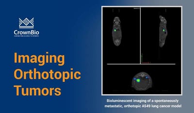
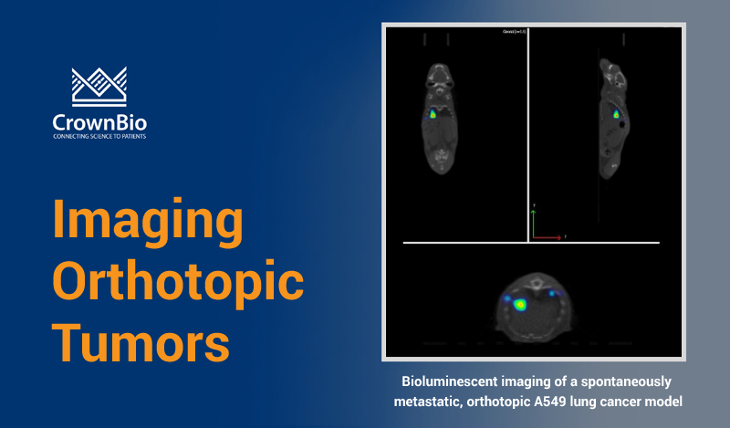
Orthotopic Imaging

Several orthotopic models have been described where cancer cells are grown at clinically relevant sites, and small animal imaging has been used to visualize disease development and response to treatment. These include models of solid tumors such as prostate, brain, lung, bone, bladder, breast, and ovarian cancer [1], as well as models of liquid disease such as leukemia.

Advantages of Imaging

As well as allowing us to visualize deep lying, metastatic, and systemic disease, preclinical imaging advantages also include the ability to predictably identify engraftment at an early stage. This allows for more expeditious therapeutic intervention than conventional models and extends the therapeutic window.

Imaging also enables us to track disease development and spread in individual animals throughout the course of a study. The presence of disease in animals can be measured before treatment starts, and at a variety of timepoints during disease development and treatment.

As each animal effectively becomes its own control, the number of animals in a scientific investigation can be substantially reduced. This complies with the principles of the 3Rs (animal replacement, refinement, and reduction) and accounts for biological variability.

The use of complementary imaging technologies allows the coregistration of anatomical data with biological data. This means we can gain a deeper understanding of the mechanisms at play. In addition, imaging also allows the visualization of appropriately-labelled entities, such as therapeutics, in real-time, in vivo. Imaging data can help to optimize dosing regimens and drug delivery strategies, refining in vivo studies and ultimately improving translation from the lab to the clinic.

Summary

Small animal preclinical imaging can provide greater insight into orthotopic models of disease. This allows us to answer a range of biological questions in an elegant, sensitive, and cost-effective manner.

  • Allowing reduction in the mouse numbers needed for a given study, with each animal followed up longitudinally numerous times throughout a study.
  • Using a variety of imaging techniques, several biological questions can be resolved in the same experiment.
  • Pharmacodynamic effects and microenvironmental factors (such as vascularity, necrosis, and apoptosis) can be examined using preclinical imaging.
  • Can be utilized across a range of disease models including cancer, neurology, CVMD, infection, and inflammation.
  • We can visualize the biodistribution in vivo of a range of novel agentsincluding antibodies, ADCs, nanoparticles, and small molecules.
  • Combining multiple imaging modalities provides biological data together with anatomical reference points.
  • A complete and comprehensive data set can be obtained, maximizing the amount of information from a study, by combining imaging with more conventional techniques such as immunohistochemistry, FACS, and PK analysis.

References

[1] de Jong, Essers, & van Weerden. Imaging preclinical tumour models: Improving translational power. Nature Reviews Cancer 2014;14(7):481–93. 

Further Reading and Information on Preclinical Imaging Techniques

PET: Kuntner and Stout. Quantitative preclinical PET imaging: opportunities and challenges. Frontiers in Physics 2014; 2 https://doi.org/10.3389/fphy.2014.00012.

SPECT: Bernsen et al. The role of preclinical SPECT in oncological and neurological research in combination with either CT or MRI. European Journal of Nuclear Medicine and Molecular Imaging 2014; 41:36-49.

MRI: Albanese et al. Preclinical Magnetic Resonance imaging and Systems Biology in Cancer Research. The American Journal of Pathology 2013;182(2):312-318.

Ultrasound: Greco et al. Ultrasound Biomicroscopy in Small Animal Research: Applications in Molecular and Preclinical Imaging. Journal of Biomedicine and Biotechnology 2012;2012:519238.

Optoacoustic imaging: Taruttis and Ntziachristos. Advances in real-time multispectral optoacoustic imaging and its applications. Nature Photonics 2015;9:219-227


Related Posts