Modern high-content imaging (HCI) and analysis platforms provide simultaneous multi-parameter visualization and quantification of thousands of cells including 3D in vitro models. This highly comprehensive information provides deep insights into the mechanisms of action, toxicity, and synergistic and off-target effects of single and combinations of compounds.
In this post, we explore the benefits of using HCI to scale up phenotypic drug discovery (PDD) strategies that allow researchers to observe and analyze drug-induced phenotypic changes using high-throughput methods.
Moving Beyond Target-Based Drug Discovery (TBDD)
Target-based drug discovery (TBDD) strategies typically involve the use of biochemical assays to evaluate binding of compounds to a known target of biological interest and ideally, disease relevance.
Historically, drug discovery was carried out using a phenotypic-based approach since the target was often unknown. However, the arrival of the genomics era led to TBDD becoming dominant as the number of ‘druggable’ molecular targets increased greatly.
Despite great successes with the TBDD approach, the focus of drug discovery efforts on single targets is thought to have contributed, in part, to high drug attrition rates. This is not entirely surprising, given that compounds often interact with multiple target proteins and pathways, producing off-target effects can negatively impact efficacy, safety and ultimately, translation to the clinic and market.
Interestingly, a comprehensive analysis showed that the contribution of phenotypic screening to the discovery of first-in-class small molecule drugs exceeded that of target-based approaches, despite the major strategy being target-based approaches. This analysis confirms that phenotypic-based approaches remain highly relevant and valuable to drug discovery efforts since they allow scientists to develop more comprehensive profiles of investigational compounds.
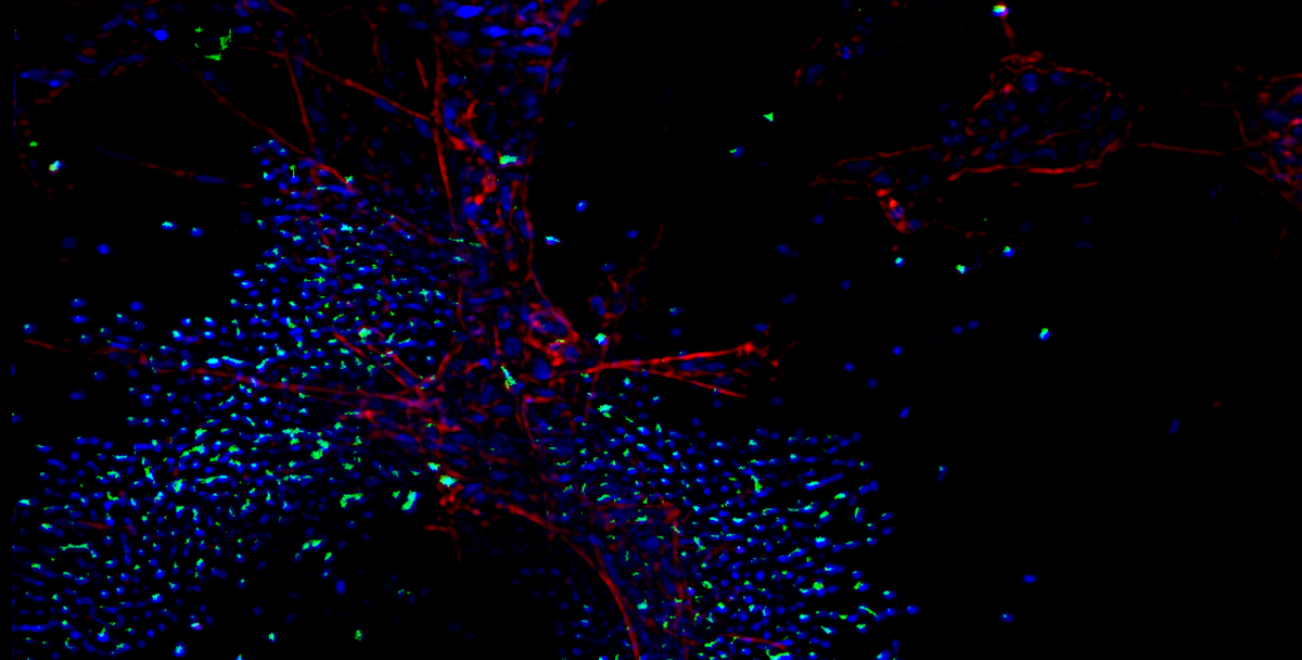
Scaling Up PDD with HCI
Cellular phenotypic changes are observable but complex traits are regulated by numerous intrinsic and extrinsic factors. With renewed interest in PDD, it is necessary to deploy technologies that enable data collection and analysis on a wide range of phenotypes in thousands of cells.
As described in a previous post, HCI broadly describes automated image-based high-throughput technology, while high-content analysis (HCA) refers to multiparameter algorithms applied to HCI data to provide detailed cellular physiology profiles within complex cell structures and microenvironments.
In short, relatively recent technological advances have introduced tools that are now being used to simultaneously assess many molecular parameters of individual cells using fluorescent dyes (including 3D organoids as described previously), including cellular and nuclear morphology, receptor internalization, cell viability, cell cycle status, protein aggregation, and more.
HCI requires specialized equipment and software designed to handle image acquisition and complex analyses. For instance, Crown Bioscience has established a customizable and scalable HCI platform that can be used to evaluate activity, toxicity, synergy, mode of action, and off-target effects of compounds.
The platform is compatible with:
384-well plates and integrated with robotic liquid handlers (e.g., Apricot S3, CyBio Felix, and Tecan Fluent)
microplate and flow cytometer-based detection systems (e.g., Tecan Infinite 200 Pro, Molecular Devices SpectraMax iD3, Luminex Guava FACS)
advanced high-content imagers (e.g., Molecular Devices ImageXpress Micro XLS, ImageXpress Micro Confocal) that can output more than 500 phenotypic changes in a single experiment.
HCI can be used to assess a multitude of drug types including antibodies, small molecules, and antibody-drug conjugates. Compounds can be applied to cells individually or in combination with readouts designed to enhance or go beyond typical fixed endpoint assays.
For example, a simple dose response experiment with HCI-generated nuclei counts can also be designed to incorporate more than 300 additional phenotypic data points to facilitate deeper analysis. Employing HCI with dose response measurements also adds visibility to efficacy panels and provides an opportunity to discover clinically relevant biomarkers. More advanced phenotypic changes can also be evaluated such as tumor and nucleus morphology, receptor internalization, protein aggregation, epithelium formation and thickness, cell polarity, swelling, and invasion, among many others.
HCI in 3D
Although high-throughput drug screening and HCI are typically associated with traditional 2D culture systems, HCI is also used with 3D in vitro models. These models, particularly patient-derived tumor organoids, offer superior clinical relevance. Unlike 2D monolayer cultures, cells placed in a 3D environment assemble into structures that resemble native tissue morphology and physiology, exhibit genetic and phenotypic stability, and carry better predictive value of patient drug response.
Similar to traditional cell lines, organoids are also amenable to long-term storage and the generation of ‘living biobanks’ has become an invaluable tool for preclinical research. Crown Bioscience’s HCI platform includes access to a large biobank of 3D cultures (e.g., classical cell line-derived spheroids, organoids, and patient tumor-derived models) with many of these accompanied by histopathology, genomic and transcriptomic data to facilitate model selection. By analyzing hundreds of phenotypic profiles in 3D models, researchers can obtain deeper insights into a compound’s effects in vitro while closely mimicking an in vivo environment, allowing for more accurate predictions of in vivo drug responses.
The value of combining HCI with 3D organoids was recently demonstrated in a recent study by Herpers et al. (2022). The researchers screened more than 500 therapeutic antibody candidates against a panel of colorectal cancer organoids and matching normal organoids. With functional evaluation provided by HCI, a novel bispecific antibody was identified that specifically triggers EGFR degradation in LGR5+ cancer stem cells, while demonstrating minimal toxicity toward healthy LGR5+ colon stem cells.
In addition to monocultures, immune organoid co-cultures can also be used with HCI to evaluate efficacy of immuno-oncology agents. For example, compounds can be tested for their ability to modulate T cell migration into tumors, immune cell priming, tumor cell killing and myeloid cell polarization. The added value of HCI is not restricted to just oncology applications. 3D culture models have also been developed for cystopathic diseases such as autosomal dominant polycystic kidney disease and cystic fibrosis. Novel 3D lumen swelling assays provide a functional readout which is easily scalable and can be used to identify on and off-target effects of potential therapeutics.
Conclusion
HCI and HCA provide researchers with simultaneous multi-parameter visualization and quantification of thousands of cells including 3D in vitro models. These technologies are being applied to PDD strategies so researchers can develop deeper insights into a compound’s effects in vitro while closely mimicking an in vivo environment when paired with 3D models, ultimately allowing for more accurate predictions of in vivo drug responses.
Learn more about our high content imaging and analysis services here.
Cite this Article
Baillargeon, M., (2022) Scaling Up Phenotypic Drug Discovery (PDD) with High-Content Imaging (HCI) - Crown Bioscience. https://blog.crownbio.com/using-high-content-imaging-for-scaling-up-phenotypic-drug-discovery
Imagine standing on the brink of a breakthrough in cancer treatment, only to be halted by one daunting barrier: drug resistance. In this episode of Cr…
Oncology drug approvals in H1 2025 In the first half of 2025, the FDA’s Center for Drug Evaluation and Research (CDER) approved a total of 16 novel dr…
Often referred to as “magic bullets” or “biological missiles”, antibody-drug conjugates (ADCs) are one of the most promising advances in targeted canc…