In this post, we explore the differences between the terms high-content imaging (HCI), high-content screening (HCS), and high-content analysis (HCA), and why these automated and image-based high-throughput technologies are increasingly being adopted to generate data to facilitate preclinical studies and downstream go/no-go decision-making.
Introduction
In large part, HCI has been enabled by the amazing advancements in microscopy that have taken place over the past few decades. Specifically, microscopes are now capable of acquiring huge numbers of images using automated processes. Briefly, HCI is an image-based high-throughput approach to cell-based screening that blends automated multicolor fluorescence imaging with quantitative data analysis to simultaneously evaluate multiple molecular features in individual cells in both 2D and 3D cell cultures, among other biological sample types. HCI is increasingly being adopted by researchers because it allows for unbiased multiparameter visualization and quantification of single cell activity across many, many cells following treatment with hundreds, thousands, or even millions of new compounds.
For instance, while a traditional low-throughput microscopy experiment may capture a biological effect (such as cell death) in a relatively small number of cells, HCI allows researchers to capture images of hundreds or thousands of cells with the desired biological endpoint. Clearly, this latter method generates a much more robust dataset since it entails capturing hundreds, thousands or even millions of images, which ultimately maximizes the number of datapoints to achieve more robust statistics.
The term “High Content” generally refers to the fact that many molecular parameters/features (which are measured using fluorescent dyes) of individual cells can be simultaneously evaluated, such as cell cycle status, cellular and nuclear morphology, cell viability, receptor internalization, protein aggregation, and so on. Thus, it is no surprise that powerful HCI-based technologies are being deployed across the spectrum of preclinical research, from identifying and validating new targets, to predicting in vivo off-target effects, among others.
While the terms “high-content imaging,” “high-content screening,” and “high-content analysis” are often used interchangeably, there are some distinctions that should be made as described in more detail below and summarized in Table 1.
The Differences Between HCI, HCS, and HCA
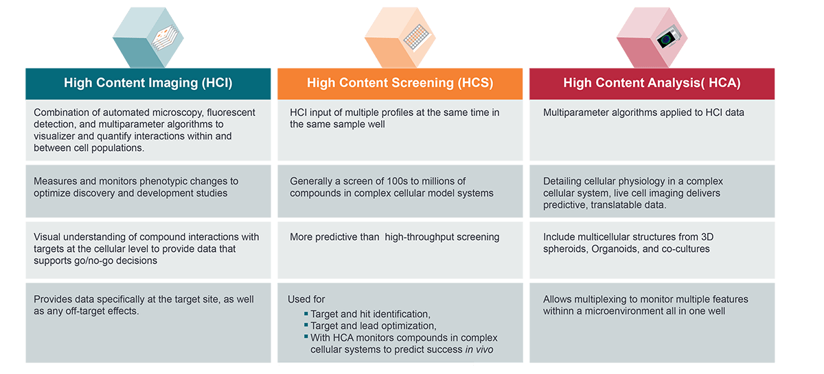
High-Content Imaging (HCI) As mentioned above, the term “HCI” is used to refer to the main underlying automated image-based high-throughput technology. HCI approaches are used to measure and monitor phenotypic changes for optimizing discovery and development studies, as well as for early discovery and high-throughput screening (HTS) to hit-to-lead and lead optimization. The ultimate goal is to develop a body of robust data that can support downstream go/no-go decisions via a better visual understanding of compound interactions with targets at the cellular level.
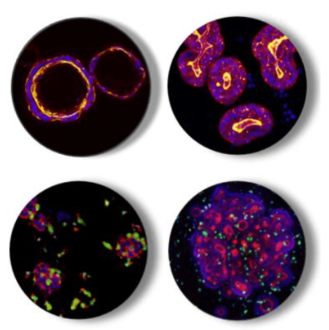
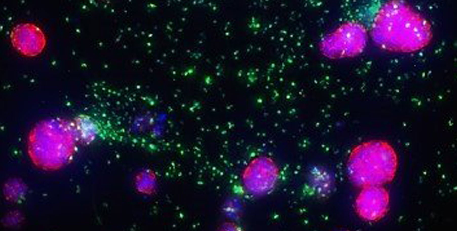
Figure 1: Representative Multiparameter HCI Images of Normal and Cancer Organoids Alone and in Immune-cell Co-cultures: Normal Colon Organoids (top left), Colorectal Cancer Organoids (top right), Tumoroids plus Myeloid Cells (bottom left), and Organoids plus Immune Cells (bottom right).
High Content Screening (HCS) The aim of HCS is akin to other traditional HTS methods that seek to screen hundreds to millions of compounds, with the objective of identifying new drug targets and hits, or target and lead optimization in complex cellular systems, including 3D cultures such as spheroids and organoids. HCS is considered more predictive than classic plate-reader-based HTS, since the HCI screening format provides phenotypic data showing the cellular response(s) to a compound, which may be related to on-target and/or off-target effects, all in the same experiment. By using multiplex techniques, multiple endpoints can be monitored, although one must take into consideration possible experimental noise, workflow challenges, and data storage capacity at the planning stages of the experiment.
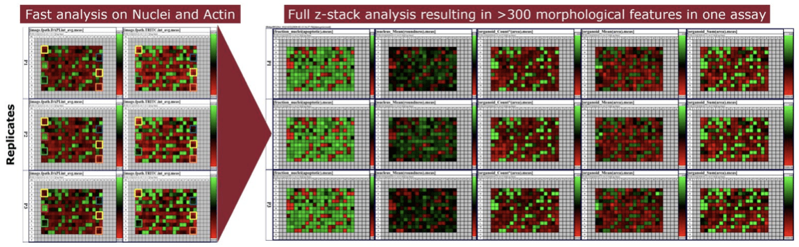
Figure 2: Example of a Representative Heatmap of HCS Data Output of Multiple Features. (Left) Fast analysis on nuclei and actin, and (Right) Full z-stack analysis resulting in more than 300 morphological assays in a single assay.
While the evolution of HCI hardware has occurred at a very rapid rate, the analytical software had not kept pace. This meant that HCS experiments were often not being used to their potential. For example, an analysis reported that despite the increasing use of HCS since 2000, the number of endpoints assessed with HCS were relatively low. The authors found that between 60-80% of published papers used only one or two endpoints, and only 5-10% used 6+ endpoints. It was suggested that this lower-than-expected use (at the time) was based on the lack of sophisticated, user-friendly, high-content analysis (HCA) software that was required for HCS experiments. Almost eight years later, it is good to know that sophisticated HCA programs have become available and are proving invaluable for analyzing multiparameter HCS data.
High Content Analysis (HCA) HCA applies multiparameter algorithms to HCS data. By developing detailed cellular physiology profiles in the context of complex cellular systems (including multicellular structures from spheroids, organoids, and co-cultures, as well microenvironments), the goal is typically to optimize hits from an HCS. In other words, sophisticated HCA algorithms are now enabling very complex analyses of cell populations in HCS experiments, ultimately delivering highly predictive and highly translatable preclinical data to predict in vivo effects.
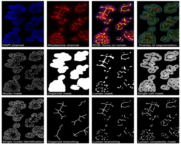
Figure 3: Example HCA Images of Organoids Used to Generate a Detailed Profile of the Complex Cellular System.
High Content Application Definitions
Screening (HCS) vs Imagin (HCI) vs Anaylsis (HCA)
Table 1: A comparison of High Content Application Definitions
Integrating High-Content Screening and Analysis with Organoids
As noted earlier, HCS can be used in conjunction with organoids, which are considered to be the most predictive in vitro model in terms of clinical response in vivo. Their remarkable clinical predictivity has to do with their recapitulation of key phenotypic (e.g., structural and cellular heterogeneity) and genetic features observed in the original tissue. In the context of oncology programs, tumor organoids have high clinical relevance and are a great platform for drug and biomarker discovery.
Conclusion
Imaged-based high-throughput technologies are enabling the development of highly predictive and highly translatable preclinical data to predict in vivo effects, especially when combined with highly patient-relevant 3D organoid cultures. While there are several challenges that must be taken into consideration prior to embarking on an HCS experiment, consulting with experts familiar with HCS instruments and workflows can provide the best opportunity for the success of your experiment.
Cite this Article
Baillargeon, M., (2021) High Content What? Deciphering between High-Content Imaging (HCI), High-Content Screening (HCS), and High-Content Analysis (HCA) - Crown Bioscience. https://blog.crownbio.com/differences-between-hci-hcs-hca
Imagine standing on the brink of a breakthrough in cancer treatment, only to be halted by one daunting barrier: drug resistance. In this episode of Cr…
Oncology drug approvals in H1 2025 In the first half of 2025, the FDA’s Center for Drug Evaluation and Research (CDER) approved a total of 16 novel dr…
Often referred to as “magic bullets” or “biological missiles”, antibody-drug conjugates (ADCs) are one of the most promising advances in targeted canc…

 In large part, HCI has been enabled by the amazing advancements in microscopy that have taken place over the past few decades. Specifically, microscopes are now capable of acquiring huge numbers of images using automated processes. Briefly, HCI is an image-based high-throughput approach to cell-based screening that blends automated multicolor fluorescence imaging with quantitative data analysis to simultaneously evaluate multiple molecular features in individual cells in both 2D and 3D cell cultures, among other biological sample types. HCI is increasingly being adopted by researchers because it allows for unbiased multiparameter visualization and quantification of single cell activity across many, many cells following treatment with hundreds, thousands, or even millions of new compounds.