3D adult stem cell (ASC)-derived tumor organoids can enhance the translatability of drug discovery data, especially in combination with high content imaging (HCI). In this blog post, we explore how the pairing of these two technologies allows researchers to develop highly comprehensive and predictive data that can enable better decisions earlier in drug discovery, while improving the translatability of the most promising lead compounds.
Current Challenges in Oncology Drug Discovery
Inter- and intra-tumor heterogeneity represents a persistent challenge in the treatment of cancer and is recognized as a major hurdle in the development of novel, safe and effective anti-cancer drugs. Despite the massive investment put forth to develop these drugs, which can take many years and billions of dollars, success has been relatively limited.
For instance, one analysis found that oncology clinical trials have a 97% rate of failure; albeit those that employ a biomarker strategy for patient-selection have higher overall success probabilities. The root cause of this statistic is due, in part, to the fact that most high throughput drug screens are conducted on 2D cell lines that are easy to establish and manipulate and can provide rapid results for early-stage decisions.
However, their physiological relevance is not ideal. For example, an analysis showed that immortalized cell lines grown in vitro and engrafted in vivo bear minimal resemblance to the tissue or tumor of origin. In addition, cell lines that have been adapted to grow outside their 3D tumor microenvironment (TME) are exposed to stressors and genetic changes that do not accurately recapitulate what occurs in patients. Thus, there is a need for more patient-relevant in vitro models that can help reduce the high rate of attrition of drugs in oncology development.
3D In Vitro Organoids for Drug Screening
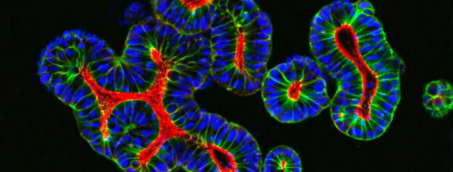
As described in more depth in previous posts (such as here and here), tumor organoids are superior in vitro 3D cultures developed using exclusive Hubrecht Organoid Technology (HUB) protocols. Building upon important discoveries in ASC biology, HUB organoids can be generated from purified single ASCs or tissue fragments that harbor resident cells with stem activity when grown under 3D conditions (e.g., intestinal crypts). Importantly, ASC-derived organoids reflect the 3D complexity found in vivo. Since the first report on the use of HUB technology to generate ‘mini-gut’ organoids, protocols have been adapted and refined to a wide variety of tissues and permit long-term expansion of organoids with maintenance through serial passaging as well as cryopreservation for future use.
Unlike other 3D in vitro systems such as spheroids, which rely on spontaneous cell aggregation, the formation of HUB organoids is driven by the unique properties of stem cells which results in many shared features with in vivo tissues such as morphology, genetic make-up and physiological functions, making them highly patient-relevant 3D in vitro tumor models for drug screening.
Tumor organoids are also highly amenable to genetic manipulation. Gene editing technologies such as RNA interference or CRISPR/Cas9 can be used to generate custom models with specific genetic perturbations that are useful in the identification and validation of biological targets and mechanistic biomarkers.
Maximizing the efficiency of 3D in vitro organoids in drug screening can be achieved by pairing it with powerful HCI technology that can penetrate the 3D structure and visualize the dynamic interplay of cells and their response to drug candidates.
The Value of Pairing 3D In Vitro Organoids with HCI
As described in a previous post, HCI generally describes automated image-based high-throughput technology and HCI and analysis (HCA) indicates multiparameter algorithms applied to HCI data. HCI and HCA platforms allow researchers to visualize and quantify the interactions of drugs with cells. Pairing patient-relevant 3D in vitro tumor organoids with innovative HCI and HCA can generate extremely detailed cellular physiology profiles of complex cellular systems, including multicellular structures from organoids and co-cultures, such as co-culturing tumor organoids with immune cells for immunotherapy efficacy studies.
HCI significantly expands the amount of information obtained from cellular assays as compared to fixed endpoint assays such as Cell-Titer Glow (CTG) or nuclei count. By combining 3D organoids with HCI early in the target/drug discovery process, the risk of downstream failure can be mitigated since only the most-promising candidates, tested on highly patient-relevant 3D models, proceed through the drug discovery pipeline. Organoid and HCI platforms are being used together for drug combination studies to inform the potential course of action for difficult-to-treat tumors that are resistant to primary treatment.
The Value of Ready-to-Use Organoid Biobanks
Commercially available tumor organoid libraries, such as our Organoid Models Database, allow researchers to access a cloud-based searchable database of patient-derived organoid (PDO) tumor models, with matched healthy organoids, for a range of cancer types, including breast, colorectal, and pancreatic, annotated with related histopathology, IC50, genomic, and transcriptomic analysis data. Biobanks capture the mutational diversity of tumors encountered by oncologists and provide a robust model to investigate disease progression, identify potential therapeutics, and study variation in drug responses produced in different patients with the same pathologies.
Further, tumor organoid biobanks are excellent platforms for screening multiple lead compounds where high-throughput screening (HTS) instruments dispense organoids into 384-well plates and introduce compounds to each well. While efficacy of lead compounds on biological targets can be monitored using traditional in vitro assays (e.g., cell viability and proliferation assays), HCI is increasingly being used to develop comprehensive data on changes in cellular response to drug candidates in high-throughput fashion.
Conclusion
Transforming drug discovery with 3D adult stem cell organoids and high content imaging aids in assessing drug toxicity, visualizing, and quantifying optimal drug concentrations, and monitoring off-target effects. These technologies allow researchers to identify biological targets, select leading drug candidates, and test drug responses in patient-relevant samples so that better decisions can be made earlier in the drug discovery process, and thus, increase the likelihood of developing a successful therapeutic.
Baillargeon, M., (2022) Transforming Drug Discovery with 3D Adult Stem Cell Organoids and High Content Imaging - Crown Bioscience. https://blog.crownbio.com/transforming-drug-discovery-with-3d-adult-stem-cell-organoids-and-high-content-imaging
Imagine standing on the brink of a breakthrough in cancer treatment, only to be halted by one daunting barrier: drug resistance. In this episode of Cr…
Oncology drug approvals in H1 2025 In the first half of 2025, the FDA’s Center for Drug Evaluation and Research (CDER) approved a total of 16 novel dr…
Often referred to as “magic bullets” or “biological missiles”, antibody-drug conjugates (ADCs) are one of the most promising advances in targeted canc…