This post explores the benefits of combining high-content imaging (HCI) and high-content analysis (HCA) with 3D in vitro tumor organoids for drug discovery and validation studies. An increasing number of large in vitro screening studies are showing that novel clinically relevant biomarkers of drug response and important drug effects can be discovered by probing a wider array of cellular parameters combined with dose–response assessments.
Why Use 3D In Vitro Tumor Organoids?
As described in a previous post, tumor organoids are highly clinically relevant 3D in vitro models available for drug discovery and validation studies. Hubrecht Organoid Technology (HUB) established robust and reliable protocols which generate tumor organoids that faithfully recapitulate original tissue physiology.
Compared to other advanced culture systems, 3D in vitro organoids are more robust and show enhanced reproducibility because they are very stable genetically and phenotypically. In addition, they have consistent growth kinetics and performance across several passages, with scalable material available for repeat studies.
In vitro 3D organoids are now more accessible due to the availability of large biobanks of well-characterized models. Such biobanks allow researchers to bring clinical trials into the lab using high-throughput platforms for screening new drugs and different drug strategies (e.g., multiple dose ratios, combination effects).
When compared to using in vivo models, tumor organoid matrix screens are highly practical for testing additive, synergistic, or antagonistic effects of combination therapies given the many possible combinations that need to be tested. Tumor organoids are value models when it comes to quantifying the efficacy and potency of oncology agents since they are readily scalable, and they are adaptable to standard in vitro assays such as IC50 measurements and CTG readouts for cell viability. Furthermore, tumor organoids are amenable to co-culturing with immune cells for early immuno-oncology efficacy drug evaluation.
Overall, tumor organoids provide a fast and cost-effective model for applications such as in vitro drug screens and demonstrate high predictivity for patient drug response. These beneficial features enable researchers to make better decisions earlier in drug discovery and increase confidence that selected drug candidates will have better translatability.
Combining Tumor Organoids with Image-Based Phenotypic Analysis
To recap from a previous post, HCI generally describes automated image-based high-throughput technology, while HCA indicates multiparameter algorithms applied to HCI data. In recent years, great technological advances have led to tools that can simultaneously assess many molecular parameters of individual cells (using fluorescent dyes), including cellular and nuclear morphology, receptor internalization, cell viability, cell cycle status, and protein aggregation.
By integrating these clinically relevant 3D in vitro tumor organoids with innovative HCA, researchers can now develop extremely detailed cellular physiology profiles in the context of complex cellular systems (including multicellular structures from spheroids, organoids, and co-cultures, as well as microenvironments). The ultimate goal of pairing these technologies is to deliver highly predictive and translatable preclinical data to define their effect on tumor organoid growth and development.
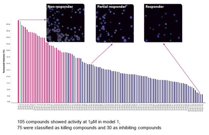
To highlight an example, patient-derived ovarian tumor organoids can be screened with a library containing thousands of compounds to define their effect on tumor organoid growth and development (Figure 1). Phase 1 would entail optimizing the experimental setup using two different ovarian tumor organoid models. This would be followed by Phase 2 where the compound library would be screened on 1-5 tumor organoid models. Phase 3, which is optional, could involve the knockdown of a gene of interest (using lentivirus for example), and this would then be followed by Phase 4 which is the identification and validation of lead compounds.
Figure 1: Thousands of compounds can be screened for lead selection using highly predictive 3D in vitro tumor organoid models.
Rich Quantitative Data for Evaluating Drug Responses
By combining a HCI screening platform with 3D in vitro organoids, researchers can measure basic parameters (such as the number, shape, and size of organoids and nuclei) and more intricate parameters (such as network formation, protrusions, lumen formation, and planar polarity). Together, these measurements can provide unique characterization of treatment effects on organoids with specific mutations and diverse genetic backgrounds. Such readout capabilities rely on advanced technologies that encompass automated liquid handling and fully integrated robotic screening platforms (e.g., Apricot S3, CyBio Felix, and Tecan Fluent), coupled with detection systems (e.g., Tecan Infinite 200 Pro, Molecular Devices SpectraMax iD3, Luminex Guava FACS), and fully automated imaging systems (e.g., Molecular Devices ImageXpress Micro XLS, ImageXpress Micro Confocal).
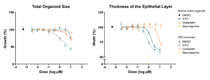
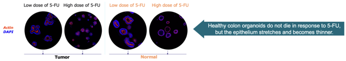
The graphs below show a study that combined HCI with patient-derived 3D tumor organoids (and matched normal organoids from the same patient) (Figure 2). The graph on the left demonstrates effective killing of tumor organoids by 5-FU. The graph on the right shows thinning of the epithelial cell layer of normal colorectal organoids. Such data can help to predict potential off-target effects in subsequent in vivo studies.
Figure 2: Representative 3D Image Analysis to Measure Tumor Killing and Nonlethal Toxicities in Tumor and Normal Organoids Derived from the Same Patient: 5-FU induced killing of tumor organoids (left) and thinning of the epithelial cell layer of normal colorectal organoids (right).
Screening and HCI Readouts
When combining a HCI screening platform with 3D organoids, there are key capabilities that the platform should offer:
Customizable 384-well plate format
Measurement of 300+ morphological parameters, including number, shape, and size of organoids and nuclei, lumen formation, and infiltration of immune cells
Quantitative analysis of growth, morphology, proliferation, cell cycle arrest apoptosis/cell death, and toxicity
Functional testing of antibodies, antibody drug conjugates, small molecules, and immune cell interactions
The following table shows the differences between traditional biochemical methods and HCI for monitoring phenotypic changes:
Monitor phenotypic changes and go beyond traditional fixed endpoint assays
Crown Bioscience Available In Vitro Applications
Biochemical
High Content Imaging
Drug Response
X
X
Drug Combinations
X
X
Cell proliferation
X
X
Cytotoxicity assays
X
X
Cell Viablility
X
X
Immuno-oncology
X
X
Tumor & Nucleus Morphology Change
X
Epithelial Changes
X
Tumor Invasion
X
Necrosis and Apoptosis markers
X
Cell Swelling
X
Cell Cycle Analysis
X
Therapy/Target Localization
X
By seeding organoids at a density of 100-500 per well (in 384-well plates), coupled with proprietary image analysis algorithms that screen, quantify, and identify the most important therapy induced changes over 300 phenotypic attributes, the combination of HCI screening platforms with 3D organoids can be used across a wide range of important applications for oncology drug discovery purposes, including the following:
Characterizing phenotypic changes in tumor organoid and immune cell co-cultures to support in vitro immunotherapy development studies
Evaluating synergistic/additive effects of combination therapies using matrix screens
Evaluating tumor organoid killing with CAR-T cell co-cultures
Assessing compounds that preferentially inhibit tumor outgrowth by screening matched normal and tumor organoid samples
Developing important insights on 3D morphological features and changes and the investigational agent’s mechanism of action
Conclusion
By pairing clinically relevant 3D in vitro tumor organoids with HCI and HCA in large screening studies, researchers can develop extremely detailed cellular profiles in the context of complex cellular systems and spatial biology. Combined with dose–response studies, this enables the discovery of new highly translatable biomarkers of drug response, further supporting downstream drug development efforts.
Combining these technologies requires very skilled and experienced scientists armed with the right tools. Contact us if you have questions on how your research program may benefit from a combination of HCI and tumor organoids.
Visit Crown Bioscience’s dedicated HCI service site to learn more about how we enable researchers to advance their drug discovery and development programs.
Cite this Article
Baillargeon, M., (2022) Pairing 3D Organoids with High-Content Imaging Can Develop Clear Views of Complex Cellular Systems - Crown Bioscience. https://blog.crownbio.com/pairing-3d-organoids-with-high-content-imaging-can-develop-clear-views-of-complex-cellular-systems
Imagine standing on the brink of a breakthrough in cancer treatment, only to be halted by one daunting barrier: drug resistance. In this episode of Cr…
Oncology drug approvals in H1 2025 In the first half of 2025, the FDA’s Center for Drug Evaluation and Research (CDER) approved a total of 16 novel dr…
Often referred to as “magic bullets” or “biological missiles”, antibody-drug conjugates (ADCs) are one of the most promising advances in targeted canc…