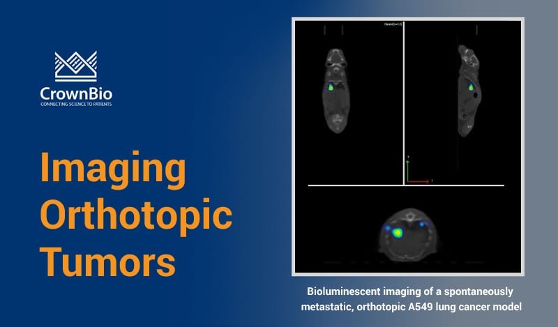
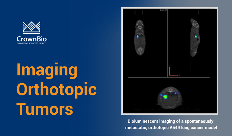
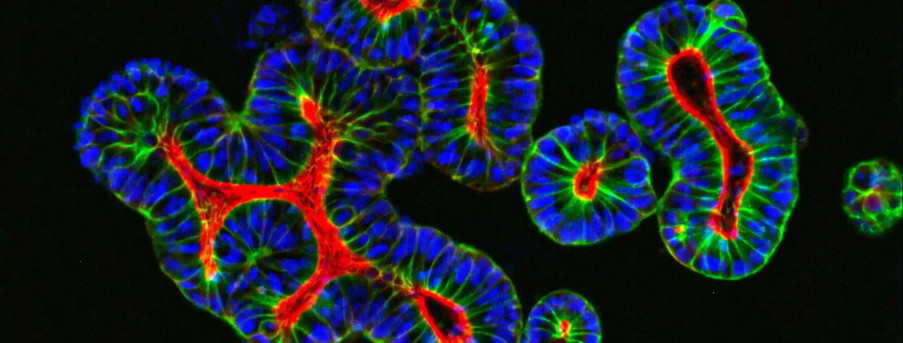
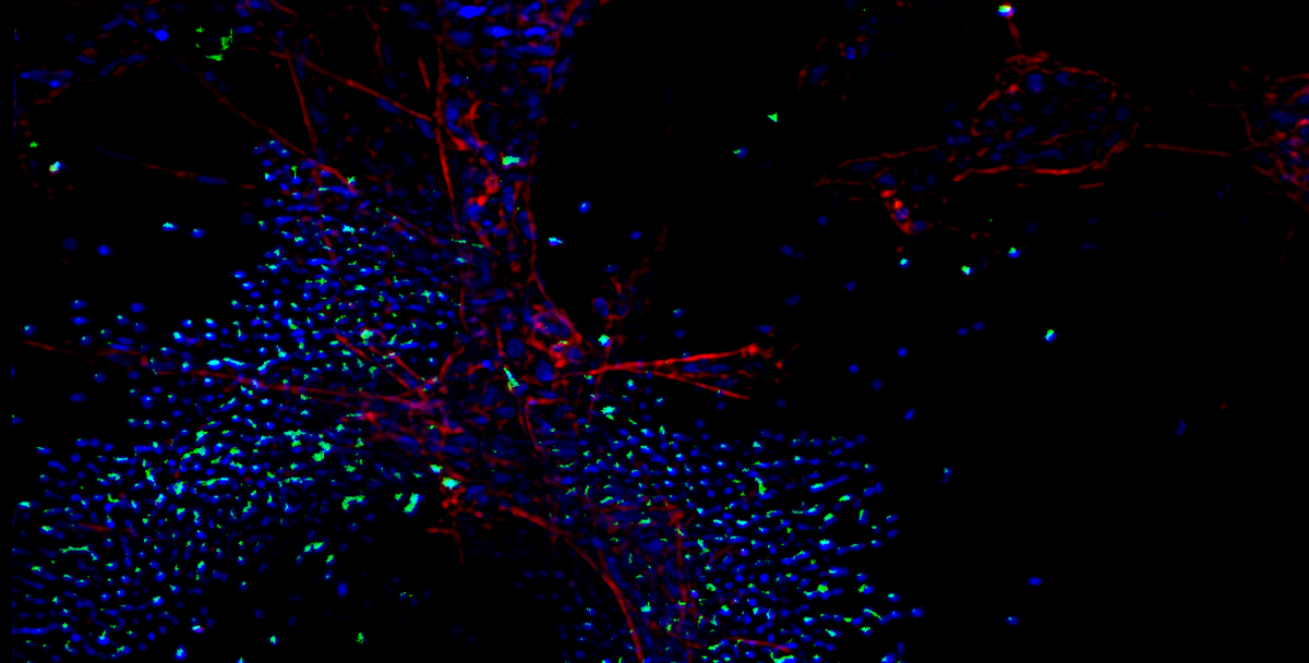
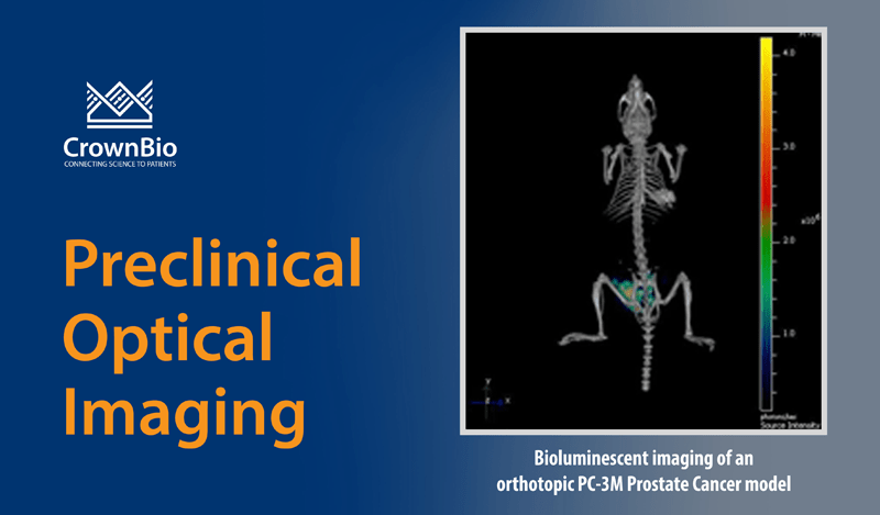
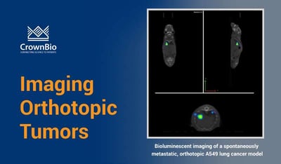
 Explore how preclinical imaging helps visualize disease grown at depth in an in vivo, orthotopic setting.
Preclinical Animal Models in Cancer Research
One of the most challenging aspects in anticancer drug research is the development of robust preclinical models to evaluate the efficacy of novel therapies. As we’ve previously discussed, subcutaneous cancer models are useful tools to help assess novel agents. However, they often don’t represent clinical disease, with differences in tumor morphology, vascularity, microenvironment, and metastatic spread.
Tumors grown at their relevant physiological site - orthotopic models - provide a more realistic environment for disease growth, and as such are valuable models for therapeutic evaluation. A major difficulty with such techniques, however, is assessment of disease burden, with deep lying, systemic, or metastatic tumors not being amenable to the usual measurement methods.
Non-Invasive Small Animal Imaging
State-of-the-art small-animal imaging modalities provide non-invasive images full of anatomical and functional information. These data make it possible to perform longitudinal studies of disease progression and response to therapy in preclinical models, including those implanted orthotopically.
Several techniques are available and may be used either individually or in combination, dependent on the biological question of interest. Small animal imaging technologies which are useful in preclinical research include:
- Positron emission tomography (PET)
- Computerized tomography (CT)
- Single Photon Emission Computed Tomography (SPECT)
- Optical imaging
- Magnetic resonance imaging (MRI)
- Ultrasound
- Optoacoustic imaging.
Each imaging modality has associated strengths and weaknesses. For example, MRI and PET/CT are relatively low throughput, and often require the use of tracers (e.g. gadolinium for MRI, 18F Fluorodexoyglucose for PET). This can increase costs and limit their use in drug efficacy studies. Optical imaging is high throughput and lower cost, but requires the use of luciferase or fluorescent markers to visualize tissues or therapeutics of interest.
Multimodal Imaging
To broaden the horizons of research, high-resolution imaging modalities are now being more commonly used in combination with sensitive functional techniques. This is across many research areas, including oncology, cardiology, infection, and neurology.
This multimodality workflow helps lead to a better understanding of underlying disease mechanisms, and provides efficient tools for evaluating new chemical entities and candidate drugs. The introduction of non-invasive small animal imaging has opened up the possibility of using such models in preclinical drug development programs. In time, this could raise the prospect of lower drug attrition rates in human trials.
Orthotopic Imaging
Several orthotopic models have been described where cancer cells are grown at clinically relevant sites, and small animal imaging has been used to visualize disease development and response to treatment. These include models of solid tumors such as prostate, brain, lung, bone, bladder, breast, and ovarian cancer [1], as well as models of liquid disease such as leukemia.
Advantages of Imaging
As well as allowing us to visualize deep lying, metastatic, and systemic disease, preclinical imaging advantages also include the ability to predictably identify engraftment at an early stage. This allows for more expeditious therapeutic intervention than conventional models and extends the therapeutic window.
Imaging also enables us to track disease development and spread in individual animals throughout the course of a study. The presence of disease in animals can be measured before treatment starts, and at a variety of timepoints during disease development and treatment.
As each animal effectively becomes its own control, the number of animals in a scientific investigation can be substantially reduced. This complies with the principles of the 3Rs (animal replacement, refinement, and reduction) and accounts for biological variability.
The use of complementary imaging technologies allows the coregistration of anatomical data with biological data. This means we can gain a deeper understanding of the mechanisms at play. In addition, imaging also allows the visualization of appropriately-labelled entities, such as therapeutics, in real-time, in vivo. Imaging data can help to optimize dosing regimens and drug delivery strategies, refining in vivo studies and ultimately improving translation from the lab to the clinic.
Summary
Small animal preclinical imaging can provide greater insight into orthotopic models of disease. This allows us to answer a range of biological questions in an elegant, sensitive, and cost-effective manner.
- Allowing reduction in the mouse numbers needed for a given study, with each animal followed up longitudinally numerous times throughout a study.
- Using a variety of imaging techniques, several biological questions can be resolved in the same experiment.
- Pharmacodynamic effects and microenvironmental factors (such as vascularity, necrosis, and apoptosis) can be examined using preclinical imaging.
- Can be utilized across a range of disease models including cancer, neurology, CVMD, infection, and inflammation.
- We can visualize the biodistribution in vivo of a range of novel agentsincluding antibodies, ADCs, nanoparticles, and small molecules.
- Combining multiple imaging modalities provides biological data together with anatomical reference points.
- A complete and comprehensive data set can be obtained, maximizing the amount of information from a study, by combining imaging with more conventional techniques such as immunohistochemistry, FACS, and PK analysis.
References
Further Reading and Information on Preclinical Imaging Techniques
Optoacoustic imaging: Taruttis and Ntziachristos. Advances in real-time multispectral optoacoustic imaging and its applications. Nature Photonics 2015;9:219-227

