In this post, we explore why and how to use in vitro screening to identify biomarkers. A case study is also provided to highlight exactly how in vitro screening can be used to develop a biomarker signature of drug response.
The Value of Biomarkers in Drug Development
Given that many diseases are heterogeneous, it is not surprising that individual patients often show widely differing responses to the same treatment.
Studies have shown that clinical trials using a biomarker strategy for patient selection are often more successful when compared to those that do not use such a strategy.
The use of biomarkers, particularly for oncology, has taken major steps forward in recent years such as when the FDA approved pembrolizumab (Keytruda®, Merck) and larotrectinib (Vitrakvi®, Bayer) to treat cancer patients based on the presence of specific tumor biomarkers, rather than the location of the tumor or site at which the cancer originated.
With the continued high failure of clinical trials, especially in oncology, there remains a significant need to identify predictive biomarkers to stratify patients to enhance efficacy while also aiming to increase patient safety.
Identifying biomarkers early in drug development can help pave the path for a robust biomarker-guided approach to identify the most promising candidate therapies at an early stage and tailor novel therapeutics for specific patient populations and indications.
Why and How to Use In Vitro Screening for Biomarker Identification?
In vitro screening studies offer a high-throughput approach for biomarker identification. This approach typically uses robust bioinformatics and validation strategies, and can leverage a range of cell types, ranging from traditional 2D cell lines to 3D organoid models (e.g., tumor organoids).
Tumor organoids are especially powerful, given that when they are established using robust HUB protocols, they have been shown to closely recapitulate the genomic, morphological, and pathophysiological characteristics of their parental tumor; providing highly clinically relevant 3D in vitro tumor models for biomarker identification.
In addition, by leveraging biobanks of patient-relevant tumor organoids which have previously been established, the heterogeneity/diversity that exists in real-world patient populations, such as cancer subtypes and genetic signatures, can be represented in vitro. This is a powerful approach when it comes to identifying biomarkers of patient drug response, for instance.
A high-throughput in vitro approach for biomarker identification starts by generating baseline profiles of the cells (e.g., genomic and/or proteomic profiles), treating them with an investigational agent, and then analyzing and comparing the profiles of responder versus non-responder samples, or correlating genomic profiles with in vitro efficacy readouts for sufficient number of cells or organoid models using bioinformatics approaches.
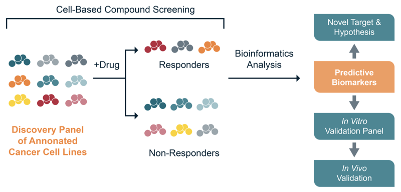
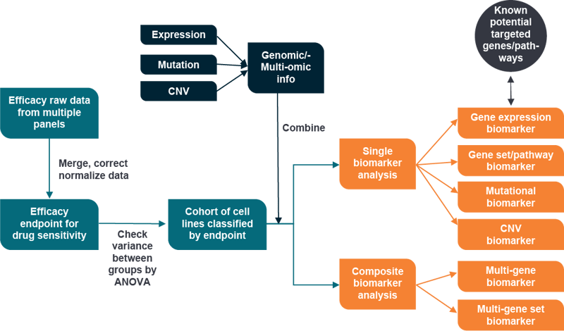
Finally, newly identified predictive biomarkers can be used to inform downstream decisions, such as the selection of appropriate in vitro and/or in vivo models for validation or novel target and hypothesis testing as shown in the image below.
The following case study highlights how an in vitro screening study can be used to effectively identify a biomarker signature of response.
Biomarker Discovery Via In Vitro Screening Case study
Irinotecan is a topoisomerase I (TOP1) inhibitor used primarily for the treatment of colon and rectal cancer. Mechanistically, irinotecan and other TOP1 inhibitors are thought to work by disrupting replication forks initiated during the S phase of the cell cycle which leads to a block in DNA synthesis, cell cycle arrest, and ultimately, cell death.
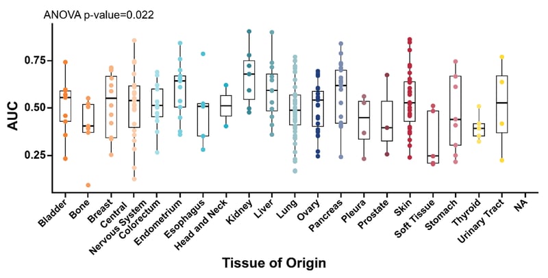
An in vitro screening study was conducted using irinotecan on a panel of 300 cell lines with the goal of identifying novel predictive biomarkers.
The following figure shows the Area Under the Curve (AUC) data obtained for the cell lines based on their tissue of origin.
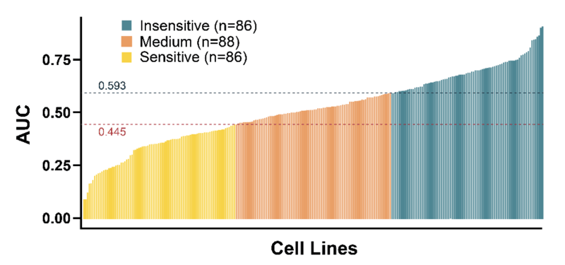
The large volume of efficacy and potency data obtained from the in vitro screen were used to classify the cell lines by irinotecan sensitivity into three groups (i.e., insensitive, medium, sensitive). This is depicted in the image below.
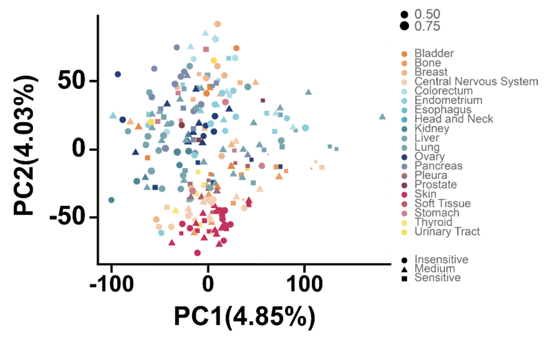
Subsequently, the global gene expression data of all cell lines was assessed via Principal Component Analysis (PCA) to identify potential ‘natural’ clustering of the groups.
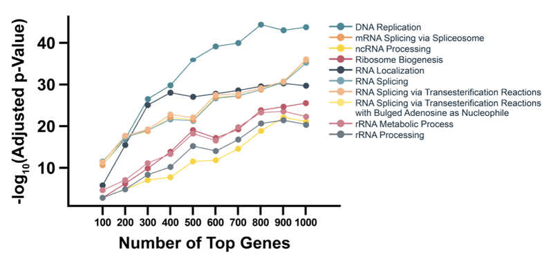
Functional analysis by Gene Ontology (GO) of Biological Processes identified DNA replication as having the most differential expression between responders and non-responders (see graph below).
This result is highly consistent with the proposed irinotecan Mechanism of Action (MoA), and also highlights the value of in vitro screens to identify and validate MoA of compounds of interest.
In Vivo Validation
A downstream in vivo validation study was then conducted using a mouse clinical trial (MCT) involving 16 unique PDX models with each PDX established in 3 to 10 mice of each treatment and vehicle arms.
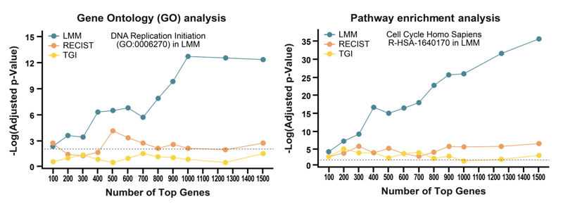
Genomic correlations with irinotecan efficacy were evaluated using a linear mixed model (LMM) framework as well as categorical endpoint-based methods (i.e., RECIST [Response Evaluation Criteria In Solid Tumors] and TGI [tumor growth inhibition]).
Consistent with the in vitro cell screen findings, bioinformatics analysis revealed that the top ranked genes were highly enriched for those involved in DNA replication initiation and the cell cycle pathway.
These data also demonstrate the importance of choosing an advance mathematical method such as Linear Mixed Model (LMM) to identify genetic correlation since it can directly model a gene’s effect on tumor growth to fit efficacy data.
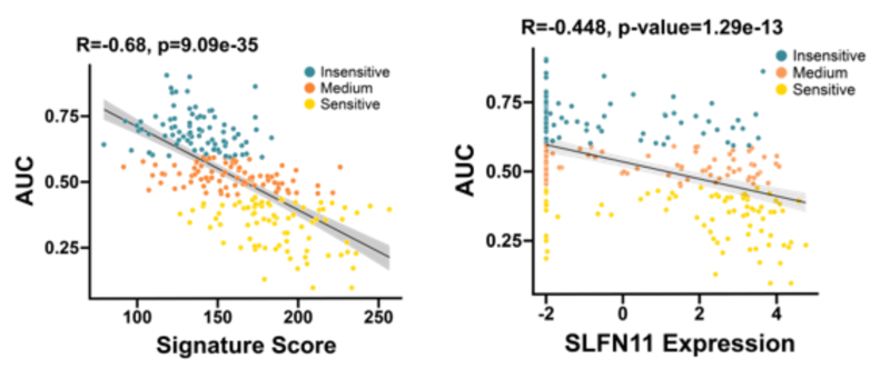
Interestingly, SLFN11 was identified as a single-gene expression biomarker of irinotecan sensitivity. SLFN11 is a putative DNA/RNA helicase that has been shown to irreversibly block replication in cells that are experiencing replication stress. Cells that express high SLFN11 have also been shown to be more susceptible to DNA-targeting drugs (see here, here, and here, for example).
However, researchers must take into consideration that a single biomarker strategy may not always be achievable, and in many cases, a single biomarker may not be ideal considering the complexities of many diseases such as cancer.
Advances in technology have opened the door to being able to measure multiple parameters simultaneously, and this approach is now leading the way for the construction of composite biomarkers that provide a more complete picture of a biological system.
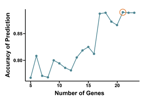
In this study, the highest prediction accuracy of irinotecan sensitivity was achieved with a set of 21 genes.
While composite biomarker panels can add complexity, it is key that downstream validation steps are carried out once a candidate gene signature is established.
As shown below, correlation between AUC and Signature Score in all cell lines, including the ‘Medium’ sensitive group, is clearly higher than AUC compared to single gene (SLFN11) expression.
To summarize the case study:
Irinotecan efficacy was evaluated across a large panel of cancer cell lines for mechanism probing, and biomarker discovery in solid tumors
A bioinformatics-driven approach to mechanism investigation showed that DNA replication is associated with efficacy via both GO analysis and pathway analysis
While expression of SLFN11 was the strongest single-gene biomarker, an expression-based 21-gene composite biomarker was able to achieve even higher efficacy prediction accuracy
For reference, the following flowchart provides a highly comprehensive in vitro biomarker discovery workflow.
Conclusion
Leveraging an in vitro screening approach for biomarker identification is a great way to incorporate a biomarker strategy early in the drug development process.
While traditional screening studies have used cell lines grown as monolayers, 3D models such as tumor organoids are increasingly being used since they have been shown to be highly clinically relevant.
As demonstrated in the above case study, single-gene biomarkers can be useful, but available technologies are now making it possible to efficiently generate composite biomarkers that have an even higher degree of accuracy.
To learn more about our in vitro screening expertise and services, please visit our biomarker discovery page.
Cite this Article
Xue, L., (2022) Biomarker Discovery via In Vitro Screening - Crown Bioscience. https://blog.crownbio.com/biomarker-discovery-via-in-vitro-screening
Advancements in oncology are increasingly driven by the ability to extract more detailed and precise information from patient tumor samples. Tradition…
Oncology continues to be a rapidly changing field with scientific discoveries transforming patient treatment and care. As it stands today, advancement…
Cancer research has evolved significantly over the years, driven by the need for more precise and efficient methods to study tumor biology, identify b…