Advanced preclinical tumor models are needed in pancreatic cancer research to develop new treatments and improve survival rates. Pancreatic ductal adenocarcinoma (PDAC), which covers up to 90% of pancreatic cancers, doesn’t respond to common anticancer drugs as well as targeted agents and immunotherapies.
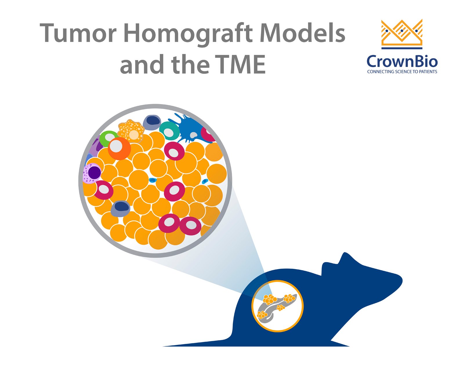
This is partially due to the tumor microenvironment (TME). The TME is made up of fibroblasts, leukocytes, and a dense fibrous stroma which is believed to shield the tumor from treatment.
In order to develop new drugs to treat pancreatic cancer, preclinical models are needed which include a human disease-relevant TME. This allows the investigation of each drugs mechanism of action and the assessment of new agent effects on the TME and tumor growth inhibition.
Tumor Homograft Models in Pancreatic Cancer Research
Conventional tumor models don’t replicate the human TME very well, but some newer, advanced models are improving on this.
Tumor homografts consist of spontaneous, carcinogen-induced, or CRISPR/Cas9-engineered GEMM tumors engrafted into immunocompetent syngeneic hosts. The models are derived from primary tumors, which are never adapted to in vitro growth, maintaining similarity to parental GEMMs. Importantly for PDAC models, tumor homografts keep relevant tumor stroma with a rich TME.
Subcutaneous vs Orthotopic Tumor Homograft Models
The TME can be affected by tumor model implantation site, which is important to consider when choosing a model for pharmacological evaluation, especially when it comes to immunotherapies that target specific immune cell populations. Subcutaneous tumors are more easily and widely used for preclinical studies. However, orthotopic models implanted in physiologically relevant sites have more biological and pharmacological relevance to human disease.
To compare implantation sites for PDAC, subcutaneous and orthotopic tumor homograft models have been developed from the same spontaneous PDAC tumor. The original tumor was from a genetically engineered KPC mouse (KrasG12D/+/P53-/-/Pdx1-cre) which captures some of the most common features of human PDAC:
Kras activating mutation
P53 loss of function.
The presence of a Pdx1-cre recombinase means these mutations are specifically induced in the pancreas, while the rest of the animal is considered wild type for these genes. This makes the original GEMM model relevant to study PDAC. As KPC GEMM are difficult to breed and not amenable to large scale studies, KPC tumor homografts represent the ideal alternative to study the anticancer effect of a new surrogate or cross-reactive agent.
A range of features were compared for the subcutaneous and orthotopic KPC homograft models, with some interesting immunoprofiling results.
Model Histology
Implantation site doesn’t affect model histology. Both subcutaneous and orthotopically transplanted PDAC mouse tumors show morphological similarity to human PDAC.
Growth Rate
Tumor homograft model growth rate is minorly affected by implantation site, with orthotopic models growing slightly quicker than their subcutaneous counterparts.
Standard of Care Response
Both subcutaneous and orthotopic models are resistant to the standard of care gemcitabine, which is to be expected based on clinical results.
Baseline Immune Profiling
The main difference between the tumor homograft model types is found when looking at baseline immune profiles. A panel of representative tumor infiltrating leukocytes (TIL) were analyzed by FACS and results compared. In both tumors, compared with a healthy pancreas, infiltrating immune cells are significantly increased. For orthotopic tumors, there’s higher B cell infiltration compared with subcutaneous counterparts.
Tumor homografts provide an advanced preclinical model for PDAC, more closely replicating the human TME for more clinically relevant studies. Both subcutaneous and orthotopic PDAC models are available and well-characterized. The main model differences lie in immune profiles, with orthotopic models showing higher B cell infiltration and increased TAM.
It’s important to consider immunoprofiling data and differences to allow the right choice when picking the most appropriate model for your preclinical efficacy study.
Imagine standing on the brink of a breakthrough in cancer treatment, only to be halted by one daunting barrier: drug resistance. In this episode of Cr…
Oncology drug approvals in H1 2025 In the first half of 2025, the FDA’s Center for Drug Evaluation and Research (CDER) approved a total of 16 novel dr…
Often referred to as “magic bullets” or “biological missiles”, antibody-drug conjugates (ADCs) are one of the most promising advances in targeted canc…