 Review the development and use of HUB Organoids, from the first discovery of Lgr5 as an adult stem cell biomarker in the Clevers lab, to the model features and data which have led to their widespread use today.
The History of Organoids
Researchers have tried to establish long term in vitro cultures of healthy human cells to study human development and morphogenesis since the late 1970s. The term ‘organoids’ became increasingly popular following the seminal works of Knoblich and Sasai. It’s currently used to signify 3D cell-based models derived from adult, embryonic, or induced pluripotent stem cells which are maintained in vitro to give rise to mini-organs.
Organoids represent the only in vitro technology to faithfully recapitulate original tissue physiology, providing a fast and cost-effective model for applications such as drug development and tissue engineering.
Adult Stem Cell Identification
It’s always been widely accepted that a pool of cells with self-renewing and differentiating properties was necessary for organ regeneration after damage. However, the identification of adult stem cells (aSCs) was elusive for a long time due to the lack of stem cell specific biomarkers.
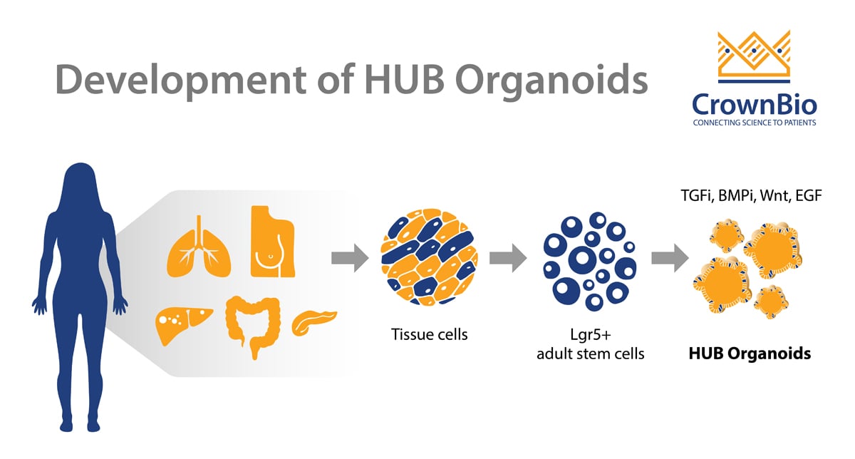
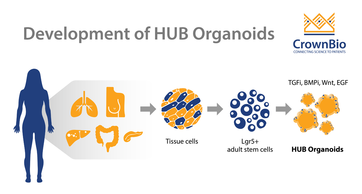
The breakthrough came with the discovery of Lgr5 as an adult stem cell biomarker for the small intestine by the Clevers lab. This finding was a steppingstone to the development of new technology mastered by Hubrecht Organoid Technology (HUB) and the large-scale production of aSC-derived organoids.
This seminal publication identified Lgr5+ cells as the stem cells of the small intestine and colon, using knock-in mouse models combined with an in-depth understanding of intestinal homeostasis and cancer regulation. Further research showed that Lgr5 is the stem cell marker in several organs of epithelial origin, such as the stomach, liver, and pancreas.
Organoid Development
Following the identification of intestinal aSCs, the Clevers lab established in vitro gut organoid cultures, starting from single Lgr5+ stem cells. Each stem cell operates independently of positional environmental cues to generate a self-organizing epithelial structure which resembles the normal gut, continuously expands, and can be cultured long term. These expanding crypt cells undergo multiple crypt fission events to generate villus-like epithelial domains containing differentiated cell types. The aSC derived organoids are remarkably stable, both genotypically and phenotypically, and can be passaged for many years.
To prove stability upon culture, a large batch of organoids was grown from a single Lgr5 colon stem cell and transplanted orthotopically into colitis models with intestinal damage. The organoids readily integrated as functional intestinal epithelium patches that were indistinguishable from the surrounding normal epithelium.
The original organoid methods were adapted to generate organoids from many different organs. The components needed to grow mini-organs using this technology are fairly conserved and include:
- a potent source of Wnt, as the Wnt pathway has emerged as the key player in maintaining the stem cell fate and driving stem cell proliferation
- a potent activator of tyrosine kinase receptor signaling such as EGF, which exerts mitogenic effects on stem cells
- inhibition of BMP/Tgfb signals which normally drive differentiation
- Matrigel to mimic the extracellular environment
Small fragments of primary tissue can also be used as the starting material for organoid development. This is potentially because tissue fragmentation and culturing in a conditioned media can mimic the environment of a damage response in vivo. This can revert cells which have already committed to differentiation back into a stem cell state.
Developing a Living Biobank of Human Tissues
Once protocols for ‘normal’ tissue organoids were established, scientists at the HUB and other research centres refined the technology to grow organoids from primary human colon, prostate, and pancreatic cancers.
HUB researchers were the first to report on the generation of a large library of colorectal cancer (CRC) tumor organoids, defined as a ‘living biobank’ of patient-derived organoids (PDOs). PDOs provide the unique opportunity for functional drug testing such as sensitivity screening and correlating treatment data with the genetic make-up of individual tumors.
HUB Tumor Organoids: Robust and Reproducible In Vitro Models
Today HUB researchers have optimized protocols for deriving and maintaining 3D ex vivo organoids from both healthy and diseased tissues, and have developed large living biobanks of PDO from several organs. HUB Organoids have emerged as a robust preclinical platform for oncology drug development, with the Clevers lab/HUB protocols widely used and published by other research groups. This is due to a number of features and applications of HUB Organoids. For example, organoid stability is a key aspect that HUB researchers have leveraged to develop applications like large scale drug screens.
HUB tumor organoids also closely recapitulate several properties of the original tumor such as gene amplification, somatic copy number, and mutations. Each organoid represents their parental patient tumor, maintaining the genetic and phenotypic characteristics. Establishment of tumor organoids from 20 consecutive colorectal carcinoma patients showed several properties of the original tumors were closely mimicked.
Genetic information is also intact in living biobanks of CRC organoids. This genetic information matches with previous large scale mutational analyses of CRC, demonstrating that CRC major molecular subtypes are represented by tumor organoids. These features mean that HUB organoids are ideal for high-throughput drug screening to investigate gene-drug associations, and to complement cell line and xenograft studies.
Phenotypic and genetic profiling of a living biobank of HUB Organoids, derived from metastatic, colorectal cancer patients showed a degree of similarity to original patient tumors. Using these organoids in drug screening showed that response or non-response to an agent is associated with organoid molecular profile.
Other studies have also compared response from ex vivo organoid, PDO-based orthotopic mouse tumor xenograft models and cancer patients in clinical trials. These data showed that PDOs faithfully recapitulate patient response in the clinic and have the potential to be used as drug screening platform.
Summary
HUB protocols provide a solid base for the development of organoids, with strong scientific background to model development. Organoid establishment allows scientists to replace 2D, regular, very homogenous cell cultures and to use the platform as a stepping stone to more expensive and time consuming animal studies. HUB tumor organoids have emerged as a robust preclinical model to predict clinical outcomes in patients, providing enhanced oncology model options for drug development programs.