It is well-known that drug attrition rates for anticancer agents are very high. In fact, only about 5% of compounds in preclinical development eventually becoming approved for clinical use. The need for more clinically relevant preclinical models has been identified as one of the areas that can reduce this attrition rate by enabling better decisions when selecting agents to advance to the clinic.
3D in vitro organoids are powerful predictive tools being used to study normal developmental processes and mechanisms of disease. Briefly, 3D in vitro organoids are microscopic self-organizing 3D structures grown from adult stem cells, and these “mini-organs” recapitulate the key structural and functional properties of the original source. Optimized HUB protocols have been established to develop models from both normal and diseased tissue, including tumor tissue (see here for a more in depth discussion on HUB tumor organoids). Based on data from numerous studies (for example, see here and here), organoids represent the first clinically relevant 3D in vitro model to show the following:
Genomic and phenotypic stability in long-term culture and after cryopreservation
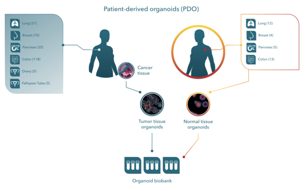
Living organoid biobanks have been developed and house many solid tumor types (with matched healthy organoids). Crown Bioscience has developed our Organoid Models Database, an extensive searchable biobank consisting of almost 350 tumor organoid models derived from its collection of PDX models (i.e., patient-derived xenograft organoids [PDXOs]) with information on histopathology, IC50, genomic, and transcriptomic analysis data associated with drug response, and an additional 200+ models from primary patient samples (i.e., patient-derived organoids [PDOs]).
These models have varied indications with multiple models derived from over 20 organ and tissue types. Cancer is a highly heterogenous disease that can differ greatly among patients with the same type of cancer (intertumor heterogeneity) and within each patient (intratumor heterogeneity). Developing effective drug candidates requires a thorough understanding of tumor heterogeneity in patient-relevant and predictive preclinical models that reflect cancer’s cellular and population-level diversity.
Biobanks capture patient population heterogeneity, support large-scale studies, and ensure availability for repeat studies. In short, biobanks bring clinical trials into the lab (referred to as “Clinical trials in a dish”) and allow researchers to identify predictive biomarkers of response (based on genomic profiling), combine drug strategies (e.g., multiple dose ratios, combination effects), and test therapeutic windows with healthy controls.
Benefits of 3D Organoids versus 2D Cell Line Cultures
Most high throughput drug screens are conducted using 2D cell lines, which are easy to establish and manipulate and are great for providing quick results for early-stage decisions. Although the physiological relevance of these cultures is not ideal, they have made significant contributions to cancer biology, and they are still very valuable when it comes to target validation and for more fundamental biological questions like defining cell signaling pathways. However, these models are suboptimal when it comes to informing about clinical response.
Increasingly, 3D in vitro organoid models are being used to bridge between traditional 2D cell lines and in vivo models. As compared to 2D cell lines, 3D organoids have a structure that promotes relevant cell–cell interactions and cell–matrix connections, possess differential areas of nutrient supply, concentration gradients of oxygen, and exposure to different environmental stressors. Together, these complex interactions and features influence a variety of cellular processes, including proliferation, drug response, signaling, differentiation, and survival. However, as compared to in vivo models, 3D organoids still lack in some of the physiologically relevant conditions provided by in vivo models.
Like 2D cell lines, 3D organoids are amenable to large-scale drug screens, and relative to in vivo models, they are more cost efficient to develop and maintain over the long term and allow for earlier decision-making instead of waiting for late-stage in vivo model data. Furthermore, since organoids preserve many of the key features of the original tumor, matched in vivo models can be generated that provide a seamless transition from in vitro studies to animal-based validation studies.
Flexible Setup for Drug Screening
Predictive 3D organoid models can be used in typical oncology-relevant in vitro drug screening assays, including:
High-throughput screens: Tumor organoids can be grown in 384-well plates for large-scale library screens, and to evaluate efficacy, toxicity, and drug combination effects. Researchers can leverage multiple organoid models, such as those derived from matched healthy tissue, different tumor types, and/or different genetic backgrounds. Engineering technologies (e.g., CRISPR, transduction) can also be used to generate models with specific features such as fluorescent labelling.
Co-cultures for immuno-therapy applications: This is a growing application of interest whereby novel immunotherapies can be tested in vitro using co-cultures of tumor organoids with various human immune cell types, including TILs, CAR-T or specific tumor microenvironment (TME) components.
Like traditional high-throughput screens with 2D cell lines, 3D organoids are adaptable to standard in vitro assays such as IC50 measurements and luminescent cell viability (e.g., CTG) readouts for cell viability. Furthermore, pairing patient-relevant 3D in vitro tumor organoids with innovative High Content Imaging (HCI) and Analysis (HCA) platforms can generate extremely detailed cellular physiology profiles of complex cellular systems, including multicellular structures from organoids and co-cultures, such as co-culturing tumor organoids with immune cells for immunotherapy efficacy studies noted above. This powerful pairing significantly expands the amount of information obtained from cellular assays as compared to fixed endpoint assays such as CTG) or nuclei count.
Deeper insights into the mechanism of a specific compound are obtained using HCI and phenotypic and morphological measurements that can be made include:
Organoid and nucleus count, size, volume, shape
Necrosis and apoptosis markers
Epithelium formation and thickness
Polarity and swelling
Internalization
Cell cycle
Therapy/target localization
IC50, area under-the-curve (AUC)
Conclusion
Cancer drug discovery and development is a costly and risky endeavor. Although simple 2D cell line cultures remain valuable in drug discovery, there is increasing interest in clinically relevant 3D organoid models that offer the scalability of 2D cell lines and the predictivity of in vivo models. 3D organoid models for cancer drug screening are a reliable and predictive tool that faithfully recapitulate the genomic, morphological, and pathophysiological characteristics of the original tumor. They are genetically and phenotypically stable even after long-term culturing and cryopreservation, and they are amenable to traditional in vitro drug screening assays and co-cultures for immuno-therapy applications.
Visit Crown Bioscience’s Organoids for Oncology Drug Development site to learn more about how help researchers advance their drug discovery and development programs.
Cite this Article
Urs, S., (2023) Organoid Technology: A Reliable Tool for Drug Screening - Crown Bioscience. https://blog.crownbio.com/organoid-technology-a-reliable-tool-for-drug-screening
Imagine standing on the brink of a breakthrough in cancer treatment, only to be halted by one daunting barrier: drug resistance. In this episode of Cr…
Oncology drug approvals in H1 2025 In the first half of 2025, the FDA’s Center for Drug Evaluation and Research (CDER) approved a total of 16 novel dr…
Often referred to as “magic bullets” or “biological missiles”, antibody-drug conjugates (ADCs) are one of the most promising advances in targeted canc…