Syngeneic tumor models are a rapid and high-throughput option for novel drug discovery. Here’s how to use them for immunotherapy target engagement studies.
Target engagement studies investigate the mechanism of action (MOA) and pharmacodynamics (PD) of a potential new drug. They are typically conducted after the feasibility of an approach has already been established through proof of concept (POC) studies.
Target engagement studies aim to identify biomarkers and correlative endpoints. These endpoints don’t represent direct clinical benefit but may help predict outcomes. Recently, there’s been an increased appreciation of correlative endpoint value, as they appear to affect the likelihood of patient response to immunotherapy.
Immunotherapy Target Engagement Studies
Due to the complex properties of the wide range of different immunotherapies, it’s become clear that they don’t fit traditional clinical trial rules. This means that novel predictive response criteria around MOA and PD need to be developed.
For example, there‘s increasing evidence that clinical trial endpoints, such as overall response rate and progression-free survival outlined by RECIST, may underestimate the benefit of immuno-oncology agents. Also, using PD-L1 expression as a biomarker has not been enough to fully explain why some patients without strong PD-L1 tumor expression show a durable clinical benefit.
The complexity of immune targeting agents lies in their nonlinear dose-response kinetics, compared to conventional therapies like chemotherapy. Immuno-oncology agents initially exert their effects by molecular target engagement either on an immune cell or a malignant cell. This then sets off immune signaling cascades that typically involve sequential steps propagating through multiple cell types (i.e. antigen presenting cells, MDSCs, T cells etc.) eventually leading to malignant cell death.
This is in stark contrast to conventional PD, where drug action is contained within a single target cell and the effect is more immediate.
Why Syngeneic Models?
In order to assess immunotherapy target engagement, a competent immune system is necessary. The use of syngeneic models enables the evaluation of MOA and PD of murine surrogate immuno-oncology agents.
To design a thorough, comprehensive target engagement study, multiple take down time points and cohorts are necessary. Syngeneic models are ideal for this purpose, as they are rapidly and cost-effectively established in a large enough scale.
How to Use Syngeneic Models for Target Engagement Studies
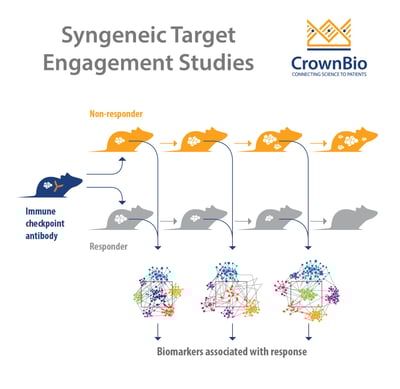
Immune and genomic profiles of treated/untreated or responder/non-responder animals are characterized to identify MOA and PD properties of a drug.
When choosing a model, it should be kept in mind that different tumor models have different baselines of number and type of infiltrating immune cells. Furthermore, the baseline may change over tumor progression.
For example, within seven days a tumor bearing model can go from a:
High level of tumor infiltrating immune cells at baseline to a lower level of immune cells overall.
Higher to lower percentage of M1 macrophages and T regulatory cells.
These factors need to be carefully considered. They can provide insight into potential predictive biomarkers based on the model response to therapy seen in proof of concept studies.
Additionally, RNAseq is used for tumor gene expression profiling to gain insights into how, and if, a specific treatment shapes tumor gene expression. RNAseq profiling also identifies key signatures of response.
Finally, immuno-PD changes upon treatment are studied in syngeneic models. Assessment of downstream effects (i.e. shifts in immune population or cytokine production) post-target engagement help to reveal an agent’s mechanism of action.
Challenges in Using Syngeneic Models for Target Engagement Studies
We’ve previously discussed that variability in tumor growth kinetics is due to the complexity of the immune system, and its tumor interaction. Unsurprisingly, there are variations among individual tumor immunophenotypes within the same treatment groups.
Similar to designing POC studies, statistics help to determine the number of animals needed for a meaningful target engagement analysis. A key point is to understand the degree of treatment efficacy variability at the POC stage. This helps to guide the number of animals (‘n’) necessary to achieve statistical significance in follow up target engagement studies. The stronger the efficacy, the less mice are needed to achieve statistical significance.
For example, if you see that all mice respond to a drug at the POC stage, then the target engagement study design is simple – evaluate the biomarker difference between control and treatment animals.
Alternatively, if you have a mix of responders and non-responders, you may want to evaluate potential biomarker differences between responders and non-responders. This therefore needs a higher n.
An important factor to keep in mind is that it’s difficult to follow the PD of individual animals. Flow cytometry is primarily a terminal analysis – meaning that the same animal is not available for subsequent time points. Biopsies are typically avoided since they may not be representative of tumor heterogeneity.
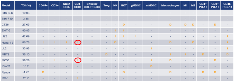
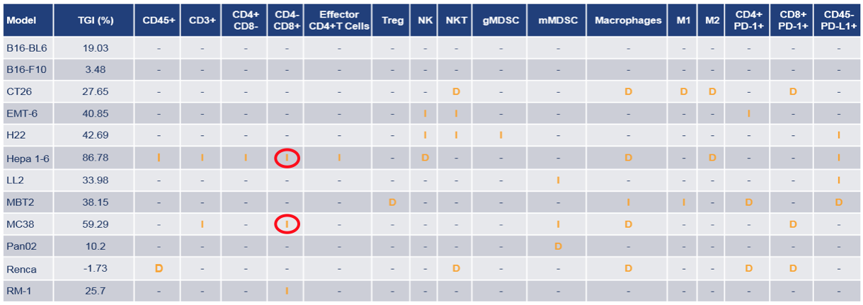
Sampling from peripheral blood can be one way to work around the limitation of terminal TIL sampling, though TIL has been more predictive in the clinical setting. Or mean values are often used to measure correlation between PD and efficacy (TGI) at a specific point in time, such as the example here.
Significant differences in mean immune cell population percentages between treatment and control group, across a panel of syngeneic models with varying mean TGI (%) at Day 13 post anti-PD-1 treatment
Another point to consider is tumor size at the time of analysis. Viability and flow cytometry gating strategies may suffer in large tumors due to necrosis. This can pose an issue for control arms (which may grow bigger than treated arms), if sampling at efficacy study terminal endpoint in the hope of correlating immune markers to response. Because of this limitation, rigorous optimization should be carried out on a model-by-model basis, and the use of sampling guidelines are recommended for target engagement studies.
Lastly, while most syngeneic tumor models offer a two to three week treatment window, this may not be sufficient to observe some immune responses. Due to non-linear response kinetics, signatures of response or any potential adverse events may manifest later, well past the allowable animal welfare endpoint criteria. Slower growing models such as Pan02 help combat this issue. Alternatively, treatment groups can be kept on study after the control animals have been taken down.
What Next?
A carefully thought out target engagement study reveals the PD and MOA of your drug and guides the inclusion of biomarkers into immunotherapy clinical trials. Integration of immunophenotyping and genomic analysis also helps identify patients more likely to respond to immunotherapies.
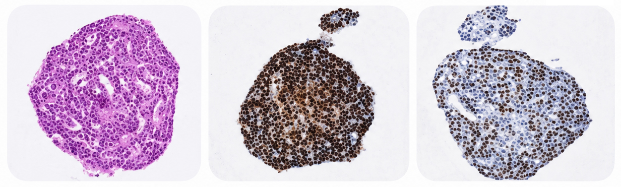
Increasing evidence suggests that a single biomarker (e.g. PD-L1) may not be sufficient. As a next step, you may want to take a meta-analysis approach to evaluate correlation between multiple immune biomarkers and response. Or perhaps you may look at the spatial relationships between biomarkers in the context of each cell’s biological role.
Ultimately, embracing the complexity of the underlying biological response to immuno-oncology agents will set you on the path to clinical success.
New data from translational research groups, platform developers, and earlystage biotech teams point to a clear trend in 2026: scalable 3D assays and …
Immunooncology (IO) drug development increasingly depends on understanding human specific immune mechanisms. However, these subtle, highly coordinated…
An Interview with Crown Biosciences' SVP of Commercial, Alex Slater Meet Alex Slater, Senior Vice President of Commercial at Crown Bioscience. With a …