Explore tumor organoid applications in cancer research including metastasis modeling, drug screening, understanding drug response/resistance and disease mechanisms, based on the unique features of these models.
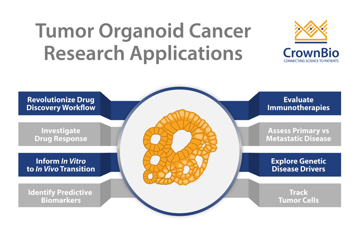
Tumor organoids have many applications in cancer research, thanks to their unique features including robustness, scalability, amenability to standard 3D assays, and translatability to in vivo engineering. These applications range from the investigation of basic disease biology, to drug response screens, through to potentially revolutionizing the traditional drug development workflow. Using tumor organoids could shift this workflow from a linear to high throughput matrix process, allowing for simultaneous selection of target patient populations and lead candidates.
These applications, among others, are discussed in this post, with uses linked to the unique features of tumor organoids.
Investigating Drug Response and Identifying Predictive Biomarkers using Tumor Organoids
Cancer is a highly heterogenous disease. This diversity is well known to oncologists, who see patient’s presenting with the same disease pathology responding very differently to the same treatment. Additionally, multiple different clones can be identified within patient tumors and between primary vs metastatic lesions, further complicating treatment response. These interpatient and intratumor variabilities are the basis for lack of response and drug resistance seen in the clinical setting.
Large biobanks of healthy and tumor organoids are used to model patient population diversity in large scale studies. Conceptually, these biobanks allow clinical trials to be brought into in vitro labs to test new treatments, refine combination strategies, and identify predictive biomarkers of response.
Organoids can also be developed from multiple regions within the same tumor. The in vitro recapitulation of intratumor heterogeneity allows both the investigation of the nature and extent of intratumor diversification, but also whether this diversity has a significant effect on drug response/resistance.
Using Tumor Organoids to Investigate Chemosensitivity and Drug Resistance Mechanisms of Primary vs Metastatic Disease
Compared to their paired primary tumor, metastatic lesions are harder to treat, often display lower chemosensitivities, and are frequently associated with drug resistance. However, the lack of highly patient-relevant in vitro models for paired primary and metastatic tumors has meant that the underlying nature of these differences remains largely unexplored.
Using tumor organoids fulfills this unmet need, since they are developed from both primary and metastatic lesions (even from the same patient) and preserve original tumor pathophysiology. Tumor organoids therefore provide a truly unique opportunity to evaluate differences in chemosensitivity and to investigate mechanisms of drug resistance.
Providing Unique Models to Understand Genetic Disease Drivers and Track Tumor Cells through Tumor Organoid Engineering
High throughput genomic technologies have allowed the identification of the most frequently mutated genes in cancer. Some of these mutations occur at a very low frequency in the patient population, and there is a shortage of models to investigate possible therapeutic interventions.
Tumor organoids can be readily engineered (more easily than in vivo models) using genome editing technologies, such as CRISPR/Cas9. This enables the knockout of specific genes to functionally validate their role in tumor progression and their role in response to treatment. Tumor organoid engineering therefore provides highly clinically-relevant preclinical models that would otherwise not exist.
Additionally, bioluminescent and fluorescent reporters can be engineered into tumor organoids to provide models that are tracked via imaging over time. This is currently possible using cell lines, however engineering patient-derived models such as PDX tumors or primary cells is technically very challenging.
Bioluminescent or fluorescent organoids are developed relatively easily compared to PDX, and can be grafted in vivo to generate new patient-relevant in vivo models. This is a particularly interesting avenue when looking at developing orthotopic xenografts to monitor tumor response to treatment over time via bioluminescent imaging.
Assessing Immunotherapies through Tumor Organoid and Immune Cell Co-Culture
In vivo testing of immunotherapies is often preferred over in vitro assessment. This is due to the complexity of recapitulating the tumor-immune cell interactions that occur in vivo and the lack of translatability from standard in vitro tumor models.
In vivo models, however, have their own challenges, such as GvHD and the partial reconstitution of human immunity in lower models.
Tumor organoids offer the flexibility of an in vitro system and can be co-cultured with various types of immune cells. Additionally, compared to traditional in vitro models they have enhanced clinical relevance.
As an alternative approach tumor organoids can be co-cultured with non-autologous immune cells to model the complex and dynamic interactions between the tumor and the immune cells in vitro.
Selecting Target Patient Populations and Lead Candidates Simultaneously Through Matrix Large Scale Tumor Organoid Drug Screens
Cancer drug discovery is a lengthy, expensive, inefficient, and linear workflow that has high attrition rates and poor translatability of current preclinical data. The current linear approach is inefficient as it involves first the identification of a lead compound, and then the selection of a target patient population.
Organoids provide a new method to move this classic linear process to a matrix workflow. In this matrix, several models (i.e. at the individual patient-level and at the patient-population level) and compounds are tested simultaneously. This speeds up the process of selecting a lead to progress and in which specific patient population.
This is made possible because tumor organoids are predictive of patient response, like in vivo PDX, but more easily scalable than any in vivo system. This makes tumor organoids particularly adaptable to large scale drug screening. Conventional and well-established in vitro assays and readouts are applied in the same way to tumor organoid screens, resulting in cleaner and higher quality data due to the robustness of the system.
Organoids are genomically stable and they can be biobanked without losing the original tissue identity and ensure availability for repeat studies. This allows for the generation of large collections of models that can be used as avatars of the patient population in preclinical drug efficacy studies.
Better Informed In Vitro to In Vivo Transition and New In Vivo Model Development
The availability of matched in vitro/in vivo models can ensure a more informed transition from early stage target identification and characterization to late-stage in vivo compound efficacy and potency validation.
However, paired models are not easy to come by, especially when looking at matched patient-derived in vitro/in vivo models.
Tumor organoids can be developed from the large collections of already established and well-characterized PDX models (PDX-derived organoids or PDXOs), providing the only clinically relevant in vitro to in vivo platform for drug testing.
Conversely, new in vivo PDX models can be developed from established organoids, effectively generating new matched pairs. This is particularly innovative when starting from engineered organoids. For example, if you use a tumor organoid engineered to express a bioluminescent probe to establish an orthotopic PDX, it becomes possible to visualize tumor response to treatment in real-time and evaluate this response in a relevant tumor environment.
Conclusion
Tumor organoid applications in cancer research are numerous and expanding due to the unique features of these models. Tumor organoids are quickly filling an unmet need for novel patient-relevant preclinical in vitro models.
Cite this Article
Barbeau, J., (2020) Tumor Organoid Applications in Cancer Research - Crown Bioscience. https://blog.crownbio.com/tumor-organoid-applications-in-cancer-research
Imagine standing on the brink of a breakthrough in cancer treatment, only to be halted by one daunting barrier: drug resistance. In this episode of Cr…
Oncology drug approvals in H1 2025 In the first half of 2025, the FDA’s Center for Drug Evaluation and Research (CDER) approved a total of 16 novel dr…
Often referred to as “magic bullets” or “biological missiles”, antibody-drug conjugates (ADCs) are one of the most promising advances in targeted canc…